Approximately one in twenty is osteoarthritis, one in ten is regularly manifested, and more than 70% of the population experiences them from time to time or singly. Problems with the musculoskeletal system are so frequent, mainly due to the irresponsible attitude to this aspect, while prevention measures require almost no special efforts.
What is it
The human musculoskeletal system is a systemically interconnected set of bones (forming the skeleton) and their joints, allowing a person to control (through impulses transmitted through the nervous system by the brain) the body, its statics and dynamics. The importance of the human musculoskeletal system is difficult to overestimate. A person whose ODS does not fulfill its functions is, at best, an invalid or a paralytic lying in a bed.
Did you know? One of the founders of anatomy in its modern, scientific form was Leonardo da Vinci. He, along with other scientists and researchers of the Renaissance, performed autopsies to understand the structure of the human body.
At healthy person ODA functions are divided into mechanical and biological.
Basic mechanical functions
Mechanical functions are associated with the preservation of the structure and movements of the body in space.
support
It consists in the formation of a basis for the rest of the body - muscles, tissues and organs are attached to the skeleton. Due to the skeleton and the muscles attached to it, a person can stand straight, his organs maintain a relatively static position relative to the axis of symmetry and each other.
Protective
Bones protect the most important internal organs from mechanical damage: the head is protected by the skull, the dorsal - by the spine, the internal organs of the chest (, lungs and others) are hidden behind the ribs, the genitals are closed by the bones of the pelvis. 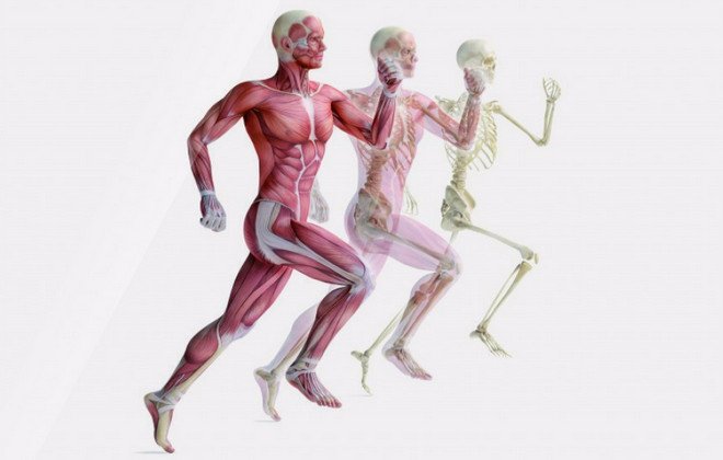 It is this protection that provides us with resistance to external influences, and well-trained muscles can enhance this effect.
Did you know? At the time of our birth, we have the most bones - 300. Subsequently, some fuse (and all become stronger) and their total number decreases to 206.
Motor
The most prominent function of the human musculoskeletal system. The creating muscles are attached to the skeleton. Due to their contractions, various movements are performed: flexion / extension of the limbs, walking and much more.
Actually, this is one of the main differences between the representatives of the biological kingdom "Animals" - conscious and controlled movements in space.
Spring
Softening (amortization) of movements due to the structure and position of bones and cartilage. 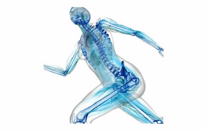 It is provided both by the shape of the bones (for example, the bend of the foot, strong tibia bones - an evolutionary mechanism that is most adapted for walking upright and supporting the weight of the body with an emphasis on only one pair of limbs), and auxiliary tissues - cartilage and articular bags provide a reduction in bone friction in their places. joints.
Biological functions of the system
The musculoskeletal system also has other functions that are important for life.
hematopoietic
The process of blood formation occurs in the so-called red bone marrow, but due to its location (in the tubular bones), this function is also referred to as the ODA.
In the red bone marrow, hematopoiesis (hematopoiesis) occurs - the creation of new blood cells, and partially immunopoiesis - the maturation of cells involved in the immune system.
Reserve
Accumulated and stored in bones a large number of substances necessary for the body, such as, and. From there they flow to other organs, where they are included in the metabolic process.  Due to these substances, the strength of bones and their resistance to external influences, as well as the rate of fusion after fractures, are ensured.
Important! Calcium problems are often caused not by insufficient calcium intake, but by rapid "washout". This is facilitated by such popular products as sweet carbonated drinks and oxalic acid. It is better to exclude all this from the diet.
Main problems and injuries of the ODA
Although the formation of the musculoskeletal system occurs in, its development is a process that continues throughout.
The causes of problems with ODA, as well as their consequences, can be different:- Incorrect load (insufficient or excessive).
- Inflammatory processes that affect bone tissue, muscles or cartilage. Depending on the etiology and localization, the diagnosis also varies.
- Metabolic disorders, deficiency or excess of any elements.
- Mechanical injuries (bruises, fractures) and the consequences of improper treatment.
Diseases of the musculoskeletal system
The diseases that affect our musculoskeletal system are depressing in their diversity:
- Arthritis affects the joints, can flow into arthrosis.
- Infections can settle in the periarticular bag (bursitis), muscles (myotitis), bone marrow (osteomyelitis), large joints (periarthritis).
- The spine may be bent, the ankle may lose tone.
Important! For any pain, see a doctor! On the early stages ODA diseases are treated with simple and sparing methods: physio- or manual therapy, therapeutic. If the disease is in a severe stage, treatment and rehabilitation will be long and difficult.
sports injuries
Of course, with proper "luck", you can fall out of the blue, and at the same time break something unexpected.
However, according to statistics, the most common injuries during sports are: muscle strains, various injuries of the lower leg, fractures (mainly the legs suffer) and ruptures (of ligaments, cartilage or tendons). 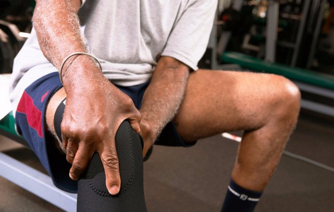
Keeping healthy: how to prevent trouble
In order to keep the body in good shape, and the ODA in a working and healthy state, it is important to know what measures to take to maintain normal functions of the musculoskeletal system.
Nothing supernatural required:
- Healthy lifestyle.
- A balanced diet rich in calcium and other minerals and trace elements.
- Regular exercise appropriate for age and health.
- Walks in the sun (vitamin D) and fresh air.
- Maintaining optimal body weight (obesity, like dystrophy, are the enemies of ODA).
- Convenient workplace.
- Regular medical checkups.
As you can see, if you support the body as a whole, everything will be in order with its systems. For this, it is not necessary to play sports professionally. 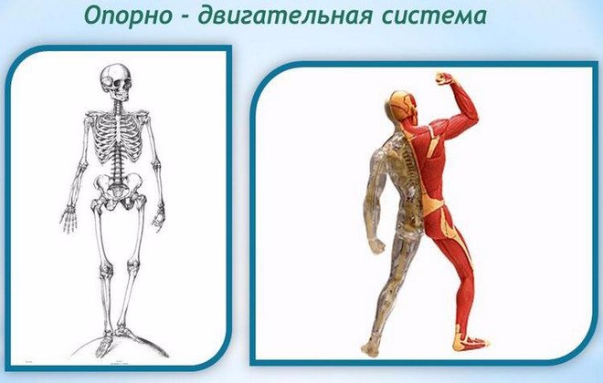 It will be enough not to neglect physical activity (in any form convenient for you, whether it be yoga, swimming or ordinary walks in the park), observe the daily routine and maintain a healthy diet. It is not so difficult. Do not be ill!
Musculoskeletal system The human body consists of a skeleton and muscles. The skeleton is the passive part of the musculoskeletal system. It is formed by bones, cartilage and ligaments. There are more than 200 bones in the human skeleton, of which 85 are paired. The human body is a collection of organs, systems and apparatuses that operate in a coordinated manner, performing vital functions. Movement is a necessary part of the function of communication and interaction, and the body can carry out this movement thanks to the musculoskeletal system. The musculoskeletal system includes bones, muscles, and bone joints. Bones are the hard and strong parts that support the body, muscles are the soft parts that cover the bones, and bone joints are the structures by which the bones are connected. All the bones, and there are about 206 of them, make up the bone system, or skeleton, which gives the body an external configuration, appearance and provides it with a rigid and durable device, protects internal organs, accumulates mineral salts and produces blood cells. Bones are composed primarily of water and minerals derived from calcium and phosphorus, and a substance called ostein. The bone is not a frozen organ: it is in a constant process of development and destruction. To do this, it has osteoblasts, bone-forming cells, and osteoclasts, cells that destroy it, so as not to allow it to thicken excessively. In the event of a fracture, osteoclasts break down bone fragments, and osteoblasts produce new bone tissue. Bone development and strength depend on the D vitamins (calciferol) that regulate calcium metabolism, which is necessary for muscle function. Especially rich in calciferol fish fat, tuna meat, milk and eggs. Also, the ultraviolet rays of the sun promote the absorption of vitamin D.
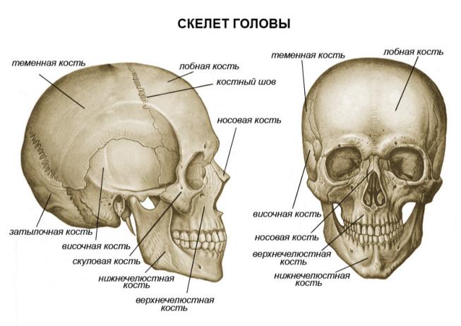
Bones facial skull - their main function is participation in chewing food.
Bones of the brain skull- the brain skull consists of eight flat bones protecting the brain, connected motionless.
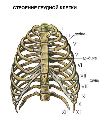
Ribs- these are the bones that, together with the sternum, form the chest, a necessary element of protection of the internal organs located in it.
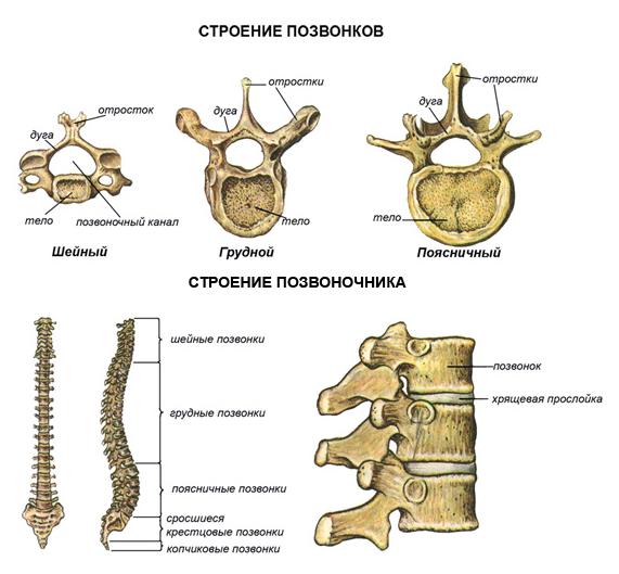
vertebral column- the axis, or support of our body, consisting of 33 or 34 vertebrae, it houses the spinal cord.
Femur is the longest bone in the human body. Allows you to make a variety of movements with your foot due to its connection with the patella.
Foot bones- a group of 26 bones, among which the largest stands out, calcaneus that forms the heel. by the most tall man there was an American in the world whose height was 2.72 m. By the time of his death, in 1940, when he was 22 years old, he was still growing. by the most low man was a 19-year-old Dutch woman: her height was only 59 cm, she died in 1895. Most long bones, about which there is information, are the bones of a brachiosaurus - a dinosaur, the remains of which were found in Colorado (USA). Its shoulder blades reached a length of 2.4 m, and some ribs exceeded 3 m. Among modern living beings, the tallest animal on Earth is the giraffe, its height can reach 6 m. only seven cervical vertebrae, the same number as a mouse. Perhaps the smallest are the temporal bones of a hummingbird - a bird whose length does not exceed 2-3 cm, but which has muscles on its wings that allow it to make up to 90 strokes per second. Hummingbirds can hover in the air when feeding on the nectar of flowers, and even fly backwards. More than 400 muscles cover the skeleton and, together with the bones and their joints, make movement possible, but some of them, such as the muscles of the veins and arteries that provide the blood flow pumped by the heart, perform functions that are not related to the motor apparatus.
Year by year, more and more aspects of life activity are revealed, on which the brain extends its supreme influence: metabolism, control of physical and chemical processes in the blood, hematopoiesis, the fight against infectious principles, etc., etc. How endlessly this is far from those ordinary-looking fibers, barely beginning to separate from the surrounding tissue, along which the primitive electrochemical excitatory impulse made its way! In higher, neokinetic animals, including us, movements are driven by sensations, controlled and directed by them. In the lower ones, on the contrary, sensations are served and provided with the help of movements. movements; apparently haphazardly and stupidly, they go ahead of sensations, grab and catch them anywhere. This mechanism of active, active "sensation" has been preserved with us, with the exception of unsystematic, in the work of our highest sense organs, vision and touch, where the circulation of the "reflex ring" is woven into a completely inseparable and very complex whole in structure. In later essays we shall have several more occasions to see with what care our central nervous system generally preserves the most ancient mechanisms, seemingly outdated and subject to archiving. This crude ancient mechanism of sensation, which operated in the most distant times, long before sensory corrections, was again revived in an improved and refined form and, merging in its work with these corrections, provided the work of our most highly developed sense organs.
Facial muscles- allow us to take on various facial expressions: laughter, anger, etc.
Biceps brachii- together with its antagonist - the triceps muscle of the shoulder - provides flexion and extension of the forearm.
External oblique abdominal muscles- allow air to be forced out of the lungs during contraction. They perform work opposite to the work of the diaphragm, which is not visible here, since it is located inside the abdominal cavity.
Quadriceps femoris- as in the case of upper limbs, the quadriceps femoris muscle also has an antagonist muscle - biceps hips. Both flex and extend the hip.
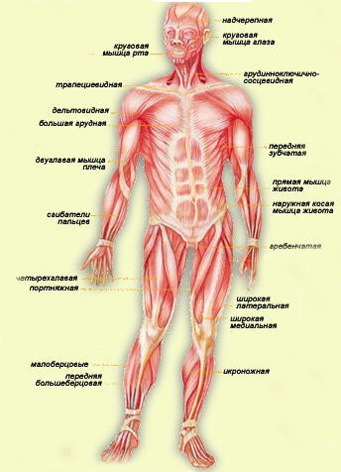
Scheme of the musculoskeletal muscular system human (front view)
Regarding sensory corrections, it should also be added that the necessary need for them, revealed in higher animals, served as a new and very powerful stimulus to further development brain. As we will show below, this need mainly contributed to the development of the so-called sensory fields, that is, whole complex casts from the sensations of the most diverse sense organs, casts that guide the movements of an animal or person and help streamline these movements in space.
Limb development
The second innovation that naturally followed the consolidation of the neokinetic system with its jointed levers and striated muscles was the development of animal limbs. The lower, skeletal organisms did not have limbs, at best, "false limbs" (pseudopodia) sometimes appeared instead of them, like the rays of a starfish or the "leg" of a snail, which, in fact, is the bottom of its body. And in vertebrates, real limbs did not develop immediately.
The limbs were a very deep, fundamental innovation. They appeared at a time when the ancient motives for the segmented (segmental) structure of the body were largely exhausted and the development of the limbs began, as it were, stepping over the ruins of this ancient principle of structure, which was still preserved on the most ancient part of the body - the trunk. Therefore, firstly, the limbs themselves no longer show any traces of segmentation - this can be seen at least in the ways in which their muscles are supplied with motor nerves. Secondly, we must point out here one circumstance that is much more important for our presentation. The successive development of neokinetics in vertebrates, followed by large motor synergies for movement in space (locomotion), and finally, limbs as improved tools for such movement, led to a corresponding enrichment of the central nervous system with adaptations necessary to serve all these evolutionary innovations. Comparative anatomy of the animal brain shows that this whole series of innovations, more than any of the previous steps of development, contributed to a real centralization in the brain, the appearance in it of the first formations, without reservations deserving of the name of the brain. The oldest part of the central nervous system of vertebrates - the spinal cord, is still fully sustained on a segmented (segmental) type of structure. The new nuclei of the brain, developed in the "fish" period of the evolution of vertebrates and finally formed in the first animal with legs - the frog, are already completely suprasegmental. Their nerve conductors already control the entire spinal cord as a whole, and in particular all the limbs. It is even more important to note the fact that the activity of this supreme brain, which controls the movements of the limbs and locomotion (we will designate it as level B in subsequent essays, proceeds in amphibians completely according to the laws of the neokinetic system: with relatively high-voltage and fast-moving electrical signals, with obedience to the law "all or nothing", etc. The more ancient centers of the brain, behind which amphibians retain control of the body (level A according to our designations), work to a large extent according to ancient motor laws: with low-voltage, slow impulses, with a large degree of participation they contain ancient, chemical signal transmission, etc. The remarkable thing here is that even we humans, the owners of a brain that is more different from the brain of a frog than a multi-story palace from a shack of a savage - even in our brains there are separate level B and level A, with decent clarity dividing the control of the limbs and the neck laryngeal muscles, and even in our country the ancient, segmental, trunk level A continues to a large extent to work according to the same ancient motor laws. We will cover the question of levels more fully in the next two essays.
Movement Enrichment
All subsequent development of movements in vertebrates is a continuous enrichment of the motor means and capabilities of animals from class to class and from "year" to "year" of our chronological table of their evolution. This enrichment does not happen without a reason and not as a result of some mysterious internal “spring” embedded in animals that impels them to continuous improvement. No, the same tough and ruthless, purely external reason leads to the enrichment of motor resources all the time: competition and the struggle for life. Animals become crowded from continuously going breeding. They don't have enough food. Predatory breeds are developed, which prefer to leave other animals to find suitable nutritional material for themselves and capture it already in a ready-made, "semi-finished" form, devouring these weaker animals. These latter develop means of self-defense: frisky legs, protective coloration, armor covers, horns and hooves, etc. Those who do not have such means of protection are first of all devoured by predators, who, without suspecting it, contribute to this improvement of the breeds pursued by them. In fact, the greatest chances of surviving extermination and producing offspring similar to themselves for a long time are those individuals who, perhaps even by chance, are better protected. And the most reliable self-defense is still rich and perfect motor capabilities. The same law of competition strikes the predators with the other end of the stick: those who are not agile, cunning and toothy among them risk dying of hunger, not being able to capture the edible living creatures that have contrived in self-defense.
Movements are enriched in this way, primarily in their strength, speed, accuracy and endurance. But this enrichment is almost only quantitative. More important are the other two sides of the movements, which are more and more improved. Firstly, those motor tasks that the animal has to solve become more and more complex and, at the same time, more and more diverse. The entire list of fish movements consists almost entirely of its main locomotion - swimming, and some pair of simple hunting movements to boot. In one of the most low-developed fish, the shark, all her hunting consists in the fact that she swims under her prey, turns her belly up (so she is more capable) and opens her mouth. In addition to swimming, an amphibious animal can also crawl, jump, make sounds. The snake already knows how to hide in ambush. And how complex and full of variety, in comparison with all this, even the chain hunting actions of a predator-mammal! Here are the tricks of the fox, and the sensitive search for a hunting dog, and the insidious ambush of the tiger, aiming at a difficult prey for him. In the next few lines, we will trace this side of the movements in more detail, the complication of the tasks they solve.
Secondly, the number of unforeseen, non-routine tasks that the animal has to solve right there, “on the go,” is increasing. As we have already seen in the introductory essay, this is where the greatest demand for dexterity takes place. In the motor life of the animal, there is relatively less and less standard, always the same movements that can be performed automatically, without delving into anything and without adapting to anything. It could be assumed that, for example, locomotion, movement in space, is an example of such, eternally patterned movements. This is far from true. When a fish swims inside a boundless aquatic environment, homogeneous in all directions, there really are not many reasons for a change. But it is a completely different matter of moving on land, which, after all, is not done on treadmills in nature. Here are ditches, and gullies, and marsh bumps, and impassable thickets; here are safe paths along which you can trot, and a forest full of secret enemies, where you need to sneak without a sound, alerting all your telereceptors, etc., etc. What can we say about more complex motor acts, completely inaccessible fish and the overflowing life of a highly developed mammal? The many times intensified struggle for life makes his existence full of surprises, and surprises require the ability to immediately, cherishing a hundredth of a second, make the right motional decision and accurately, deftly implement it. We will see further that this unceasing increase in the number of unlearned movements and actions is based on the same unstoppable development of completely new, higher parts of the brain, mainly the so-called cerebral cortex.
The first rudiments of the cerebral cortex appear already in higher reptiles, but only in higher vertebrates - in mammals - does it seize a decisive predominance and continuously develop further and further. It is the cerebral cortex that is a brain organ that has an unlimited ability to absorb the personal life experience of an animal, remember it, master it with meaning and create on its basis one-time solutions to new, previously unseen problems. In terms of mental activity, this ability is quick wit, sharpness, reason; in terms of motor acts, we call this same ability dexterity. It is not for nothing that they often say about a person endowed with pronounced dexterity: “What smart movements he has! What smart hands.” The brain matures in a human baby floor by floor in the same order in which they arose in the animal world. - the floor-level of the pallidum B, which is just finishing its development - the "ceiling" level of amphibians. Therefore, the child is not able to make any movements that would go beyond the limits of the meager list of this level. The matter is further complicated by the fact that the more ancient and lower located level A , which will be discussed below and which controls the movements and positions of the neck and trunk, does not have time to mature and enter into operation by the time of birth.Because of this, it turns out, first of all, that the newborn cannot control the main support of the whole body - the trunk and neck, holding his head, and therefore unable to use his "dynamic props" - limbs.His torso lies helplessly on his back, heavy and motionless, and all four paws can make only erratic kicking movements in all directions idly. And besides this, there is another complication: level-floor B, as already mentioned, has access for its impulses to motor praklets spinal cord, and through them - to the muscles only as a "transit", through the nuclei of the underlying level A. Therefore, he himself is forced to wait in inactivity until level A finally matures and begins to pass through his motor impulses. This deprives the child of the synergy that level B brings with it - coordinated integral movements of the limbs, and even more so the joint work of all limbs. Practically speaking, during the first two or three months after birth, there is no motor coordination whatsoever. It is only towards the end of the first quarter of life that the correct joint movements eyes, turns from the back to the stomach, etc. Near the end of the first half of the year, more or less simultaneously come into operation: the lowest level A, which gives the baby a harmonious and strengthened body, and the level of the striatum (CI), which enables him to sit, get up on your feet, stand, then crawl on all fours (again, a biogenetic memory of our four-legged ancestors!) and, finally, walk and run. The pyramidal system of the cortex (pds) lags even more. The sensitive sections of the cortex come into operation much earlier: the child begins to recognize what he sees, and understand the words addressed to him, and find a sense in the taste, gastronomic sensations. PDS begins to gradually manifest itself during the second half of the year, following the striatum system. This is reflected in the fact that the child learns to grasp what he sees in front of him, to put and shift things, to point with his finger, etc. The first monosyllabic meaningful speech sounds, usually demonstrative and pleading (like " give!"). The movements of the handles are still very inaccurate, the child often and grossly misses, but until that time he did not even attempt to make such movements as grasping or throwing. He had nothing to do with them! The difference between infants after and before six months in regard to these movements is about the same order of magnitude as the difference between the owner of a bicycle, who still barely knows how to ride it, and a person who does not have a bicycle at all. Thus, the aggravation of the struggle for existence gradually accumulated more and more significant numbers of motor tasks that are homogeneous among themselves, so far unbearable for animals. The need to cope with them grew with increasing inevitability over time. The animal had to satisfy these increasingly complex motor needs at all costs if it did not want to die. And on the way to such satisfaction there was one obstacle, the main and main one: the need to master new sensory corrections.
Consists of a skeleton and muscles, it performs the following functions:
Protective (limits the cavities in which the internal organs are located);
support function;
Provides active human movements;
Performs a hematopoietic function;
Participates in metabolism.
The passive part of the musculoskeletal system is the skeleton, consisting of bones, cartilage, joints and ligaments. There are over 200 bones in the human skeleton.
Each bone is an organ bone tissue.
Bone\u003d cells with processes + intercellular substance + nerves + vessels + connective tissue membrane
Bones:
(properties of bone): organic substances (flexibility and elasticity), inorganic substances (hardness).
Direction of growth (source of new cells): in length (cartilage), in thickness (periosteum).
Connection of bones: movable, semi-movable, fixed
Joint– articular bone with articular cavity + articular bone with head + strong ligaments + articular bag + joint fluid
Human skeleton consists of 200 bones.
Main departments:
muscles- the active part of the musculoskeletal system, providing all the variety of movements performed in the human body. Thanks to the muscles, the body maintains balance, moves in space, respiratory movements are carried out by the chest and diaphragm, swallowing, a voice is formed, eye movements are carried out, the work of internal organs, including the heart. There are two types of muscles in humans: smooth and striated.
Smooth muscles are found in internal organs: the walls of blood vessels, Bladder, ureters, intestines. Their reduction occurs arbitrarily.
Striated muscles provide attachment of muscles to the tendons and bones of the skeleton. Skeletal muscles move the bones relative to each other in the compositions, in addition, they are involved in the formation of the walls of the abdominal and chest cavities, the pelvis. They are part of the wall of the upper part of the esophagus and larynx. Carry out the movement of the apple, respiratory and swallowing movements. All skeletal muscles can be divided into two groups - flexors and extensors.
Mimic muscles are the muscles of the face, not connected with the joints.
The heart muscle is a special striated muscle, where the fibers are connected, it contracts rapidly.
In humans, each muscle contains all types of muscle fibers; their ratio varies depending on the purpose of each muscle. Blood vessels approach each muscle, which penetrate the outer shell and break up in the muscle into a network of capillaries. Muscle fibers are supplied with oxygen and nutrients through the blood. In addition, each muscle has a nerve that transmits signals.
The organs of movement are a single system, where each part and organ is formed and functions in constant interaction with each other. The elements that make up the system of organs of movement are divided into two main categories: passive (bones, ligaments and joints) and active elements of the organs of movement (muscles).
The size and shape of the human body is largely determined by the structural basis - the skeleton. The skeleton provides support and protection for the entire body and individual organs. The skeleton has a system of movably articulated levers, set in motion by muscles, due to which various movements of the body and its parts in space are performed. Separate parts of the skeleton serve not only as a container for vital organs, but also provide their protection. For example, a skull rib cage and the pelvis serve to protect the brain, lungs, heart, intestines, etc.
Until recently, the prevailing opinion was that the role of the skeleton in the human body is limited to the function of supporting the body and participating in movement (this was the reason for the emergence of the term " musculoskeletal system"). Thanks to modern research, the understanding of the functions of the skeleton has expanded significantly. For example, the skeleton is actively involved in metabolism, namely in maintaining at a certain level mineral composition blood. Substances included in the skeleton, such as calcium, phosphorus, citric acid and others, if necessary, easily enter into exchange reactions. The function of the muscles is also not limited to the inclusion of bones in movement and the performance of work, many muscles, surrounding the body cavities, protect the internal organs.
General information about the skeleton. Bone shape
The human skeleton is similar in structure to the skeleton of higher animals, but has a number of features that are associated with upright posture, movement on two limbs, high development of the hand and brain.
The human skeleton is a system consisting of 206 bones, of which 85 are paired and 36 unpaired. Bones are the organs of the body. The weight of the skeleton in a man is approximately 18% of the body weight, in a woman - 16%, in a newborn - 14%. The skeleton consists of bones of various sizes and shapes.
According to the shape of the bones are divided into:
but) long (located in the skeleton of the limbs);
b) short (located in the wrist and tarsus, i.e., where greater strength and mobility of the skeleton are simultaneously required);
in) wide or flat (form the walls of the cavities in which the internal organs are located - hip bone, bones of the brain skull);
G) mixed (have a different shape).
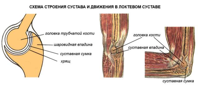
Bone joints
Bones articulate in a variety of ways. According to the degree of mobility, joints are distinguished: a) fixed; b) sedentary; c) movable joints of bones, or joints.
An immovable joint is formed as a result of the fusion of bones, while movements may be extremely limited or completely absent. For example, the immobility of the bones of the brain skull is ensured by the fact that numerous protrusions of one bone enter the corresponding recess of the other. This connection of bones is called a suture.
The presence of elastic cartilage pads between the bones provides little mobility. For example, such pads are available between individual vertebrae. During muscle contraction, the pads are compressed, and the vertebrae are drawn together. During active movements (walking, running, jumping), cartilage acts as a shock absorber, thereby softening sharp shocks and protecting the body from shaking.
Movable joints of bones are more common, which is provided by the joints. The ends of the bones that form the joint are covered with hyaline cartilage 0.2 to 0.6 mm thick. This cartilage is very elastic, has a smooth shiny surface, so the friction between the bones is significantly reduced, which greatly facilitates their movement.
From a very dense connective tissue, an articular bag (capsule) is formed, which surrounds the articulation area of \u200b\u200bthe bones. A strong outer (fibrous) layer of the capsule firmly connects the articulating bones. Inside the capsule is lined with a synovial membrane. The joint cavity contains synovial fluid, which acts as a lubricant and also helps to reduce friction.
Outside, the joint is reinforced with ligaments. A number of joints are strengthened by ligaments and inside. In addition, inside the joints there are special devices that increase the articulated surfaces: lips, discs, menisci from connective tissue and cartilage.
The joint cavity is hermetically closed. The pressure between the articular surfaces is always negative (less than atmospheric), and therefore the external atmospheric pressure prevents their divergence.
Types of joints
According to the shape of the articular surface and along the axes of rotation, the joints are distinguished:
but) with three;
b) with two;
in) with one axis of rotation.
The first group consists of spherical joints - the most mobile (for example, the joint between the scapula and humerus). The joint between the innominate and the thigh, called the walnut, is a type of ball and socket joint.
The second group is elliptical (for example, the joint between the skull and the first cervical vertebra) and saddle joints (for example, the joint between metacarpal bone first finger and corresponding wrist bone).
The third group includes block-shaped (joints between the phalanges of the fingers), cylindrical (between the ulnar and radius bones) and helical joints (forming the elbow joint).
Any loose body has six degrees of freedom, because it produces three translational and three rotational movements along the coordinate axes. A fixed body can only perform rotations. Since all links of the body are fixed, joints with three axes of rotation are the most mobile and have three degrees of freedom. Joints with two axes of rotation are less mobile, therefore they have two degrees of freedom. One degree of freedom, which means that joints with one axis of rotation have the least mobility.
The structure of the bone
Each bone is a complex organ consisting of bone tissue, periosteum, bone marrow, blood and lymphatic vessels and nerves. With the exception of the connecting surfaces, the entire bone is covered with periosteum - a thin connective tissue sheath rich in nerves and blood vessels that penetrate from it into the bone through special openings. Ligaments and muscles are attached to the periosteum. The cells that make up the inner layer of the periosteum grow and multiply, which ensures the growth of the bone in thickness, and in the event of a fracture, the formation of a callus.
Sawing a tubular bone along its long axis, one can see that a dense (or compact) bone substance is located on the surface, and under it (in depth) - spongy. In short bones, such as vertebrae, spongy matter predominates. Depending on the load experienced by the bone, the compact substance forms a layer of different thickness. The spongy substance is formed by very thin bony crossbars oriented parallel to the lines of the main stresses. This allows the bone to withstand significant loads.
The dense layer of bone has a lamellar structure and is similar to a system of cylinders inserted into each other, which also gives the bone strength and lightness. Bone tissue cells lie between the plates of bone substance. Bone plates make up the intercellular substance of bone tissue.
A tubular bone consists of a body (diaphysis) and two ends (epiphyses). On the epiphyses are located articular surfaces, which are covered with cartilage involved in the formation of the joint. On the surface of the bones are tubercles, tubercles, grooves, ridges, notches, to which the tendons of the muscles are attached, as well as holes through which the vessels and nerves pass.
The chemical composition of the bone
Dried and defatted bone has the following composition: organic matter - 30%; minerals - 60%; water - 10%.
The organic substances of the bone include fibrous protein (collagen), carbohydrates and many enzymes.
Bone minerals are represented by salts of calcium, phosphorus, magnesium and many trace elements (such as aluminum, fluorine, manganese, lead, strontium, uranium, cobalt, iron, molybdenum, etc.). The skeleton of an adult contains about 1200 g of calcium, 530 g of phosphorus, 11 g of magnesium, i.e. 99% of all calcium present in the human body is contained in the bones.
In children, organic substances predominate in the bone tissue, so their skeleton is more flexible, elastic, easily deformed during prolonged and heavy load or incorrect body positions. The amount of minerals in the bones increases with age, and therefore the bones become more fragile and more likely to break.
Organic and mineral substances make the bone strong, hard and elastic. The strength of the bone is also ensured by its structure, the location of the bone crossbars of the spongy substance in accordance with the direction of pressure and tension forces.
Bone is 30 times harder than brick and 2.5 times harder than granite. Bone is stronger than oak. It is nine times stronger than lead and almost as strong as cast iron. In a vertical position, the human femur can withstand the pressure of a load of up to 1500 kg, and the tibia - up to 1800 kg.
Development of the skeletal system in childhood and adolescence
During fetal development in children, the skeleton consists of cartilage tissue. Ossification points appear after 7–8 weeks. The newborn has a ossified diaphysis tubular bones. After birth, the ossification process continues. The timing of the appearance of ossification points and the end of ossification are different for different bones. Moreover, for each bone they are relatively constant; they can be used to judge the normal development of the skeleton in children and their age.
The skeleton of a child differs from the skeleton of an adult in its size, proportions, structure and chemical composition. The development of the skeleton in children determines the development of the body (for example, the musculature develops more slowly than the skeleton grows).
There are two ways of bone development.
1. Primary ossification, when bones develop directly from the embryonic connective tissue - mesenchyme (bones of the cranial vault, facial part, partly the clavicle, etc.). First, a skeletal mesenchymal syncytium is formed. Cells are laid in it - osteoblasts, which turn into bone cells - osteocytes, and fibrils impregnated with calcium salts and turn into bone plates. Thus, bone develops from connective tissue.
2. Secondary ossification, when the bones are initially laid down in the form of dense mesenchymal formations that have the approximate outlines of future bones, then turn into cartilaginous tissues and are replaced by bone tissues (bones of the base of the skull, trunk and limbs).
With secondary ossification, the development of bone tissue occurs by replacement both outside and inside. Outside, the formation of bone substance occurs by osteoblasts of the periosteum. Inside, ossification begins with the formation of ossification nuclei, gradually the cartilage resolves and is replaced by bone. As the bone grows, it is resorbed from the inside by special cells called osteoclasts. The growth of bone substance comes from the outside. Bone growth in length occurs due to the formation of bone substance in the cartilage located between the epiphysis and diaphysis. These cartilages are gradually shifted towards the epiphysis.
Many bones in the human body are not formed entirely, but in separate parts, which then merge into a single bone. For example, the pelvic bone initially consists of three parts, merging together by the age of 14–16. The tubular bones are also laid in three main parts (ossification nuclei in the places where bone protrusions are formed are not taken into account). For example, the tibia in the embryo initially consists of a continuous hyaline cartilage. Ossification begins in the middle part at about the eighth week of intrauterine life. Replacement on the bone of the diaphysis occurs gradually and goes first from the outside, and then from the inside. At the same time, the epiphyses remain cartilaginous. The nucleus of ossification in the upper epiphysis appears after birth, and in the lower epiphysis - in the second year of life. In the middle part of the epiphyses, the bone first grows from the inside, then from the outside, as a result of which two layers of epiphyseal cartilage remain separating the diaphysis from the epiphyses.
In the upper epiphysis of the femur, the formation of bone trabeculae occurs at the age of 4–5 years. After 7–8 years, they lengthen and become uniform and compact. The thickness of the epiphyseal cartilage by the age of 17–18 reaches 2–2.5 mm. By the age of 24, the growth of the upper end of the bone ends and the upper epiphysis fuses with the diaphysis. The lower epiphysis grows to the diaphysis even earlier - by the age of 22. With the end of ossification of tubular bones, their growth in length stops.
Ossification process
The general ossification of tubular bones is completed by the end of puberty: in women - by 17-21, in men - by 19-24 years. Because men reach puberty later than women, they tend to be taller on average.
From five months to one and a half years, that is, when the child gets on his feet, the main development of the lamellar bone occurs. By the age of 2.5–3 years, the remnants of coarse fibrous tissue are already absent, although during the second year of life, most of the bone tissue has a lamellar structure.
Decreased function of the endocrine glands (anterior pituitary, thyroid, parathyroid, thymus, genital) and lack of vitamins (especially vitamin D) can cause delayed ossification. Acceleration of ossification occurs with premature puberty, increased function of the anterior part of the adenohypophysis, thyroid gland and the adrenal cortex. Delay and acceleration of ossification most often appear before the age of 17–18, and the difference between “bone” and passport ages can reach 5–10 years. Sometimes ossification occurs faster or slower on one side of the body than on the other.
With age, the chemical composition of bones changes. The bones of children contain more organic matter and less inorganic matter. With growth, the amount of salts of calcium, phosphorus, magnesium and other elements increases significantly, the ratio between them changes. So, in young children, calcium is retained in the bones the most, but as they grow older, there is a shift towards greater retention of phosphorus. Inorganic substances in the composition of the bones of a newborn make up one-half of the bone weight, in an adult - four-fifths.
A change in the structure and chemical composition of bones also entails a change in their physical properties. In children, the bones are more elastic and less brittle than in adults. Cartilage in children is also more plastic.
Age-related differences in the structure and composition of bones are especially pronounced in the number, location, and structure of the Haversian canals. With age, their number decreases, and the location and structure change. How older child, the more dense matter in his bones, in young children there is more spongy substance. By the age of 7, the structure of tubular bones is similar to that of an adult, however, between 10–12 years, the spongy substance of bones changes even more intensively, its structure stabilizes by 18–20 years.
How younger child, the more the periosteum is fused with the bone. The final demarcation between bone and periosteum occurs by the age of 7. By the age of 12, the dense substance of the bone has an almost homogeneous structure, by the age of 15, single areas of resorption of the dense substance completely disappear, and by the age of 17, large osteocytes predominate in it.
From 7 to 10 years of age, the growth of the medullary cavity in the tubular bones slows down sharply, and it is finally formed from 11–12 to 18 years of age. The increase in the bone marrow canal occurs in parallel with the uniform growth of the dense substance.
Between the plates of the spongy substance and in the medullary canal is located Bone marrow. Due to the large number blood vessels in the tissues of newborns there is only red bone marrow - hematopoiesis occurs in it. From six months, a gradual process of replacing the tubular bones in the diaphysis of the red bone marrow with yellow, consisting mostly of fat cells, begins. Red brain replacement is completed by 12–15 years of age. In adults, red bone marrow is stored in the epiphyses of tubular bones, in the sternum, ribs and spine and is approximately 1500 cubic meters. cm.
The healing of fractures and the formation of callus in children occurs after 21-25 days, in infants this process occurs even faster. Dislocations in children under 10 years of age are rare due to the high extensibility of the ligamentous apparatus.