Recently, the number of biceps tendon ruptures has become more frequent. This is due to the great popularity of bodybuilding, football and snowboarding. Young people tend to get this kind of injury due to falls and old injuries. People over 40 can also get torn biceps due to tendon wear and certain shoulder problems. Since such an injury carries many unpleasant consequences (limitation of movement, pain, decreased strength when bending in the shoulder, etc.), we are in a hurry to tell you about how the rupture of the biceps tendon is treated.
A ruptured bicep can happen anywhere. A disease such as a complete and partial rupture of the biceps tendons can occur due to many factors. But there are a few main ones. First, this chronic inflammation, which often cause microtraumas, which lead to ruptures. Secondly, athletes often experience overexertion due to high loads on the biceps tendon, which also causes injury. Thirdly, if the rotator cuff is damaged, rupture may also occur.
Another cause of a ruptured biceps muscle is taking certain medications (statins), which cause the tendons to tear from the bone. Often, chronic ulnar bursitis is accompanied by irritation, which causes the tendon to detach from the radius.
Symptoms of a torn bicep:
- Spherical swelling on the anterior surface of the shoulder. If you relax the biceps, it will disappear;
- Sharp pain accompanied by a clicking sound;
- Subcutaneous hemorrhages.
In addition, in some people, this injury is asymptomatic. They may feel tightness between the elbow and shoulder. This indicates chronic damage to the biceps tendon.
Diagnosis of biceps tendon rupture.
In order to accurately diagnose damage to the biceps, it is necessary to undergo an examination by a traumatologist. Most often this is enough to make a diagnosis. But it is best to use instrumental diagnostic methods. To do this, you can use X-ray, ultrasound or MRI. Of course, the most informative method is magnetic resonance imaging. With its help, you can find out even the smallest changes in the structures of the shoulder
Treatment of a torn biceps tendon.
Used to treat young patients surgical methods treatment. Tenodesis is performed - an operation during which the tendon is fixed with sutures to the fascia or bone near the joint, or subacromial decompression. Doctors are trying to use arthroscopic techniques.

But for people of middle and old age there is conservative treatment rupture of the biceps muscle. Such therapy is meant by unloading and exercise therapy. But most often, doctors use an individual comprehensive treatment strategy, which provides for treatment taking into account the characteristics of the person.
To reduce inflammatory processes use drug treatment rupture of the bicep. The drugs also have an analgesic effect, which is especially important during the acute phase of the injury.
After a biceps tendon rupture, athletes are allowed to return to sports no earlier than 4 months later. At this time, it is physiotherapy, which helps people recover from a torn bicep.
MUSCLE RUPTURESIn athletes, damage to contracted muscles most often occurs, i.e. muscles that are at the peak of the contractile phase, and there is almost no damage in the relaxation phase. Muscle injuries are usually closed, i.e. no damage skin. Open muscle injuries occur much less frequently and are not difficult to diagnose. They are found and sutured during the primary surgical treatment of the wound. Closed muscle tears may be complete or incomplete. The term "muscle strain" used to be used, but now it is practically not used, since it is believed that in any case there are ruptures of individual fibers. muscle tissue. Closed muscle tears occur with a sharp, unexpected movement of the muscles, or a reflex contraction as a defensive reaction (fall).
Muscles unprepared for the load, not warmed up, or, on the contrary, very tired, are more likely to succumb to ruptures. The localization of muscle ruptures to a large extent depends on the sport. Weightlifting, throwing, volleyball, all kinds of wrestling, rowing, skiing lead to a rupture of the trapezius muscle.
The long back muscles are prone to tearing in those involved in weightlifting, rowing, throwing, skiing (slalom), and diving. These same sports, as well as gymnastics, wrestling, volleyball, basketball, can contribute to muscle tears. shoulder girdle: deltoid muscle of the shoulder, supraspinatus muscle. Rowing, gymnastics, weightlifting, wrestling are characterized by ruptures of the biceps of the shoulder. Triceps shoulder is damaged in those involved in volleyball, throwing, weightlifting, in water divers. Quite often there are ruptures of the quadriceps muscle of the shoulder.
Sports: jumping, sprinting, hurdling, diving, weightlifting. Other thigh muscles - extensors are prone to tearing in runners, gymnasts, wrestlers. The adductors of the thigh are often torn in football players, slalomists, volleyball players, and basketball players. Calf muscle susceptible to damage in runners, jumpers, gymnasts, wrestlers, skiers.
Treatment of muscle tears. Cold should be applied immediately after breaking. Then anesthesia is given. Small partial muscle tears are treated conservatively. In athletes, to fully restore muscle function, muscle tears of more than 25% of the muscle thickness must be treated promptly.
If a partial rupture of the muscle is treated conservatively, then a plaster cast is applied to the limb, in the position of muscle relaxation. Carry out physiotherapy and recommend a dosed load. For large and complete muscle tears, treatment is surgical. The muscle is sutured with U-shaped sutures. Sports traumatologists use plastic surgery using pieces of femoral fascia or hard meninges, as it is believed that the suture material cuts through the muscle tissue, and the use of fascia allows.
MUSCLE HERNIATION
A muscle hernia occurs when the fascia of that muscle is torn as a result of an injury. Fascia is a connective tissue sheath that covers the muscle. The most common cause of rupture of the fascia and the formation of a muscle hernia is a direct blow to the muscle. Such gaps also occur during the start of runners. Occasionally, muscle hernias occur after surgical interventions. Muscle bundles bulge through the torn fascia and, when the muscle contracts, a protrusion in the form of a hemisphere is formed. When probing, it is elastic, compacted during muscle contraction.
Muscular hernias usually occur on the thigh, with a rupture of the wide fascia of the thigh, and on the lower leg. In ordinary people, quite often such hernias do not disrupt vital activity and do not require treatment. In athletes, due to the high requirements for muscle function, treatment is almost always surgical. Often it is difficult to simply sew the fascia, or the seam is fragile. In these cases, resort to plastic surgery, the type and volume of which depends on the qualifications of the surgeon and the presence of grafts.
migeloses
migeloses called painful seals in the form of nodules in the muscle. It is believed that seals in the muscle occur due to improper loads. In weightlifters, volleyball players, tennis players, handball players, such nodules occur in the muscles of the shoulder girdle. And in those athletes who load the lower limbs more (running, jumping, football), migeloses occur in the muscles of the legs, pelvic girdle. In the rest, most often painful seals occur in the region of the trapezius muscle, back muscles.
For the prevention of migelosis, the muscles must be fully warmed up before a heavy load, and after the load, massage should be performed. The same massage is also used to treat migelosis, and the massage can be painful. Physiotherapy is used.
breaks distal tendon biceps brachii (biceps).
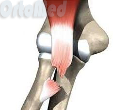
The biceps brachii, or biceps, is located on the front surface of the shoulder. She bends her hand elbow joint and, to some extent, provides outward rotation of the forearm (supination). In addition, the biceps brachii plays important role in stabilization shoulder joint, i.e. prevents dislocations. The muscle consists of two heads, which are attached to the scapula with two separate tendons at the top, and below the muscle with a single tendon (distal tendon) is attached to the radius. You can read more about the anatomy of the biceps brachii here (click to go to another article).If there is a complete rupture of the distal tendon, then the biceps of the shoulder is displaced upward. In this case, the tendon simply cannot grow to the place on the radius to which it is normally attached.
The frequency of ruptures of the distal tendon of the biceps brachii is 1-2 cases per 100,000 population per year.
In addition to rupture of the distal tendon, the tendons of the long or short heads of the biceps brachii can also rupture.
1 – elbow bone, 2 - humerus, 3 - radius, 4 - distal tendon of the biceps of the shoulder, 5 - tendon of the short head of the biceps of the shoulder (in the illustration, this head deceptively looks like a long one), 6 - scapula, 7 - coracoid process of the scapula, 8 - acromial process of the scapula, 9 - long head of the biceps brachii and its tendon (10).
Why does the break happen?
The most common distal biceps tendon tear occurs in men older than 35 when they carry or lift something heavy with their arms in front of them (for example, when they carry a heavy box in front of them). Lifting weights, especially with a jerk and without taking into account its weight, is a vivid example of such a situation. Breaks in women are extremely rare.Unfortunately, with age, in some people, the tendons lose their strength, and in cases where the mass of the object being carried or lifted is more than critical, a rupture may occur. You can prevent a gap by performing a warm-up before significant physical effort, but, unfortunately, this simple requirement is often neglected. However, a man doing a warm-up before bringing a box of food from the car home would look very unusual, you see. It is worth noting that regular physical education, and not occasionally, will be able to strengthen the tendon tissue. No other effective ways there is no way to strengthen the tendons (except maybe prolotherapy, which is in the process of clinical research). A variety of biological additives also do not have real effectiveness, and recommendations for eating plenty of jellied meat, meat broths, chicken cartilage are nothing more than common myths.
Additional risk factors for distal biceps tendon ruptures include:
Smoking: nicotine can interfere with the nutrition of tendon tissue.
Introduction of corticosteroids. Administration and local injections of corticosteroids (drugs such as diprospan, hydrocortisone) can cause tendon necrosis and rupture.
In addition, there are suggestions that some systemic diseases and taking fluoroquinolone antibiotics.
What happens during a break?Biceps tendon tears can be partial or complete.
Partial breaks. As the name suggests, these tears are incomplete, and because part of the tendon remains intact, the muscle does not move upward.
Complete breaks. This type of tear is much more common than a partial tear. A complete rupture means that the muscle is completely detached from the bone and is pulled by its contraction to the shoulder joint. Distal biceps tendon ruptures are equally common in both right hand, and on the left.
It is important to know that even with a complete rupture of the distal tendon, the patient can independently bend the arm at the elbow. This is possible due to the fact that not only the biceps muscle is responsible for flexion, but, for example, also the shoulder and muscle.
If the integrity of the distal biceps tendon is not restored, then flexion at the elbow will continue to be provided only by the brachialis muscle, which provides 60-65% of the total flexion force at the elbow. The strength of supination of the forearm will decrease to a greater extent with a bent arm in the elbow joint. In many cases, this is enough for everyday life, but, unfortunately, in order to bring purchases from the car home, you will have to do about twice as many trips, and serious sports physical activity will be completely impossible.
Symptoms
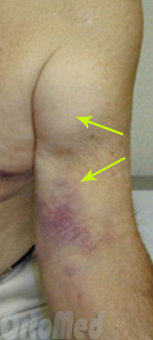
At the moment of rupture, a “click” is often felt in the elbow area. As we have already noted, with ruptures of the distal tendon of the biceps biceps muscle, the muscle is retracted (shifted) upward and its abdomen takes the form of a ball. Bruising in the area of the elbow joint is also often observed.
Immediately after the rupture, the pain is quite pronounced, but then it gradually disappears and almost completely disappears a couple of weeks after the injury.
Other symptoms are:
Edema on the anterior surface of the elbow joint.
Visible bruising in the area of the elbow joint and forearm. A few days after the rupture, the area of the bruise becomes larger, it gradually descends and may even reach the hand.
Weakness when bending the arm in the elbow joint.
Weakness when rotating the forearm (supination).
A ball-shaped seal in the upper part of the shoulder, formed by a contracted muscle.
Falling down on the anterior surface of the elbow joint as a result of the absence of the tendon.Breaks and breaks big chest muscle
Tears and avulsions of the pectoralis major muscle are a fairly rare injury that occurs, as a rule, in sports people and in the vast majority of cases in men. More common are complete detachments of the muscle, or rather its tendon from the place of attachment to humerus, but there are also partial breaks. Surgery is required to fully recover from a complete rupture. In this article, you will learn why pectoralis major tears occur, what they are, and how pectoralis major tears are diagnosed and treated. In addition, we will talk about rehabilitation after ruptures of the pectoralis major muscle.
Anatomy
The pectoralis major muscle refers to the muscles of the chest or the muscles of the girdle upper limb. In addition to it, the muscles of the girdle of the upper limb include the deltoid, supraspinatus, infraspinatus, large and small round, subscapular and some others.
The pectoralis major muscle has three parts or bundles: the clavicle, sternocostal and abdominal (abdominal). The clavicular part starts from the lower part of the inner half of the clavicle, the sternocostal part starts from the sternum and from the cartilages of the five upper ribs, respectively, and, finally, the smallest abdominal or abdominal part starts from the sheath of the rectus abdominis muscle.
All three parts of the pectoralis major fuse together to form one short, flat tendon that attaches to the humerus just below the greater tubercle.
Attachment of the pectoralis major muscle to the humerusCauses of rupture of the pectoralis major muscle.
In the vast majority of cases, ruptures of the pectoralis major muscle occur in male athletes (amateurs and professionals) at the age of 20-50 years. As a rule, a rupture occurs when the muscle is overstretched (when performing a chest press, which is the cause of a rupture in about half of the cases) or when hitting the arm at a time when the pectoralis major muscle is tense (in volleyball, when turning over in kayakers, arm wrestlers, when playing rugby, etc.).
It is known that rupture of the pectoralis major muscle is facilitated by the use of steroid drugs and a number of diseases (systemic lupus erythematosus, etc.), but in many cases the rupture occurs in people who do not have any diseases and have never used steroid drugs.
What are pectoralis major ruptures?
breaks pectoralis major muscle is considered a rare injury. But in fact, the true frequency of ruptures is unknown. The fact is that people with ruptures do not always go to the doctor, and if they do, then in the vast majority of cases these are cases of a complete rupture, but there are also partial ruptures that are perceived by the athlete as "sprains" and with such "trifle" injuries remain unknown, but in fact this may not be so rare.
Partial break can occur anywhere in the pectoralis major muscle: in the muscle itself, at the point of transition of the muscle into the tendon (muscle-tendon junction), in the tendon itself, at the point of attachment of the tendon to the humerus.
In most cases, partial tears are treated as "sprains", or the athlete does not go to the doctor at all, as he is sure that he only "pulled his hand." Luckily, partial tears heal perfectly and there is no trace of the injury, but sometimes, after trying to return to training, a partial tear causes pain, and then you have to do an operation. In the world scientific medical literature, isolated cases of operations with partial ruptures are described, so it is impossible to draw any scientifically based conclusions about the best tactics for treating partial ruptures.
There are 5 types of ruptures of the pectoralis major muscle:Type 1: Avulsion of the tendon from its attachment to the humerus (the most common type of rupture, mostly complete ruptures, rarely partial).
Type 2: A tear where the muscle joins the tendon (usually partial, not complete).
Type 3: rupture of the muscle fibers themselves (rare, may be complete or partial).
Type 4: Avulsion of the tendon of the pectoralis major muscle with a bone block (avulsion fracture). It is extremely rare.
Type 5: Avulsion from attachment to sternum, ribs. It is extremely rare.Symptoms
At the time of injury, sometimes you can even hear the sound of crackling, tearing. Immediately after this, there is a sharp pain in the chest, shoulder and the location of the pain depends on where the rupture occurred. The pain may "shoot" down the arm. After a few tens of minutes, a bruise appears in the shoulder area, which is a classic proof of a rupture - after all, there are muscles and tendons inside blood vessels and when a muscle is ruptured, these vessels also rupture. As a result, a hemorrhage occurs, a hematoma is obtained and the blood permeates the subcutaneous fat, skin. Later, this bruise travels down the arm over the course of several weeks, gradually brightens and disappears.
At full break the muscle contracts and crawls away to the middle of the body, and at the point of rupture it becomes visible to the west. Since the torn muscle cannot fully perform its function, weakness occurs in those movements for which this muscle is responsible. large muscle(pushing from the chest, bringing the arm to the body, etc.). Later, as the decline acute pain from the break itself, strength is gradually restored, but at the expense of other muscles. Unfortunately, with a complete rupture, self-recovery of strength is never complete: if you do not perform an operation with a complete rupture, then only half of the strength is restored!
For partial breaks there is no retraction, and the bruise may be small or completely absent. This causes certain difficulties in diagnosis.
Diagnostics
The doctor must determine the type of tear. Sometimes the diagnosis can be made during a routine examination, and in doubtful cases, the doctor may prescribe an ultrasound, MRI or X-ray. However, these research methods often erroneous results and we have repeatedly had to deal with the fact that, for example, ultrasound gives a picture of a partial rupture of the muscle part, but in fact, during the operation, we find a complete detachment from the attachment site. This is due to the fact that ultrasound and MRI specialists very rarely see such an injury and therefore often make mistakes in their conclusions.
Treatment
When the tendon of the pectoralis major muscle is torn from its attachment to the humerus (type 1 tear), surgery is preferable. Studies have shown that without surgery, strength is restored by only 56% and there is often pain, and surgery allows you to return 90-97% of strength and reduce the frequency and severity of painful manifestations. With such gaps after the operation, even a return to big sport is possible. Ideally, the operation should be performed as early as possible: after all, a non-working pectoralis major muscle, with a complete rupture, will inevitably atrophy and weaken.
Currently, so-called anchor fixators are used to fix the tendon of the pectoralis major muscle to the bone. These are special devices that have a special latch at one end, to which very strong threads are attached. According to the type of material from which the fixative (anchor) itself is made, they are of two types - absorbable and non-absorbable. Non-absorbable fixators are metal (usually made of titanium alloys), they are made in the form of a screw that is inserted into the bone channel and remains there forever. In general, modern alloys are very safe and a long stay of the retainer does not cause any problems. The advantage of non-absorbable (metal) retainers is that they are more durable. Another version of the fixative is absorbable. We believe that it is better to use non-absorbable fixators to fix the detached pectoralis major muscle.
The essence of the operation is that the tendon is attached to the place on the humerus from which it came off. The difficulty lies in the fact that it is impossible to simply sew the tendon to the bone - the bone is hard. Therefore, earlier one or two screws were screwed into the bone, to the heads of which the torn tendon was tied with strong surgical threads.Type 1 breaks(rupture of the tendon of the pectoralis major muscle) can be operated on even many years after the injury. Of course, the results of the operation with chronic ruptures (and ruptures older than three months are considered chronic) are worse compared to those cases when the operation was done quickly, but still it allows you to improve strength and reduce the frequency and severity of pain.
The complexity of the operation in the case of an old rupture may lie in the fact that the muscle that has been in a contracted state for a long time may not be able to be pulled to the site of attachment, and in this case, plastic surgery may be required by using a tendon from another place (tendon hamstring, etc.) , due to which the missing length is restored. The need for this is extremely rare. It is believed that if before the operation the pectoralis major muscle is in a contracted state inside from the nipple, then there is a high probability that plastic surgery will be required.
It is important to note that in rare cases, pain may persist after the operation, and such an insulting result is not always due to a mistake - the operation can be done technically perfectly. Fortunately, in the vast majority of cases, the operation gives positive results.
Type 2 tear treatment(a rupture at the point of transition of the muscle into the tendon) is debatable. Some surgeons prefer to operate on such tears immediately, while others find it worthwhile to wait a few weeks until a scar forms at the site of the tear. The fact is that the tendon of the pectoralis major muscle is small and it is technically difficult to sew a detached muscle directly to the tendon. Moreover, expectant management with type 2 rupture is more justified with partial ruptures, when after scar fusion there may not be any problems in the form of a decrease in strength or pain, and, accordingly, there will be no need for surgery.
Type 3 breaks(rupture of the muscle fibers themselves) is usually technically impossible to stitch and most surgeons prefer to treat such tears without surgery. In rare cases, with a significant size of such a gap, the operation is still performed, but for stitching the muscle, this requires a plastic material that strengthens the place of the gap (another tendon, for example, from the leg, or fascia).
Gap Type 4(rupture of the tendon of the pectoralis major muscle with a bone block or avulsion fracture) is treated with surgery. The detached piece of the humerus is fixed with a screw and / or the anchor fixators already described by us are used, with the threads of which the tendon is stitched.
Type 5 breaks(muscle detachment from the sternum, ribs) are so rare that there are no generally accepted recommendations and the decision on the method of treatment is made in each case individually.
It can be noted that type 1 ruptures are very favorable for surgery - the vast majority of patients regain strength and almost completely get rid of pain, while surgery is possible even many years after the rupture. If there is a partial tear, especially type 2 or 2, and the patient himself has low physical requirements, then it is quite possible to do without surgery.
Usually, for sewing on a detached pectoralis major muscle, an incision is made 10-15 centimeters long, which is quite traumatic.
Complications
Complications after surgery are rare. It is important to understand that the stitched tendon can also come off, and this problem can occur when training is resumed.
How can a tear/tear of the pectoralis major muscle be prevented?
When bench pressing at the very beginning of lifting the barbell and when lowering the barbell to the chest, the hands on the bar should not be too wide apart - it is in this position that the load on the pectoralis major muscle can be prohibitive. In addition, do not lower the bar of the bar directly to the chest.
Rehabilitation
Postoperative rehabilitation program after suturing the pectoralis major muscle
(Please note that the timing of certain exercises will be determined only by your attending physician, the presented program is purely advisory in nature)Goals and Exercises
I phase of rehabilitation: 1-14 days after surgery
Treatment of pain, inflammation, swelling and surrounding tissues Cryotherapy (local application of cold), non-steroidal anti-inflammatory drugs (painkillers), rest for the hand.
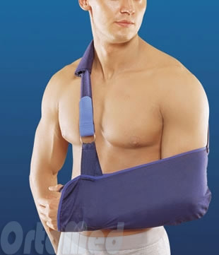
Stitched muscle protection Hand rests on a gusset or sling. You also need to sleep in a sling bandage. Nothing can be lifted by hand. You can’t take your hand to the side, forward, you should avoid sudden movements, falls, you can’t lean your hand on the railing, etc. Do not put on socks with a operated hand, etc. Outerwear is thrown over the arm.
Prevention of contractures of the shoulder and elbow joint Once or twice a day, the arm at the elbow must be completely bent and extended (from 5-7 days after the operation), and light rocking and circular motions with an amplitude of 10-15 degrees.
II phase of rehabilitation: 3-5 weeks after surgery
If necessary- treatment of pain, inflammation, swelling of the joint and surrounding tissues
The need rarely arises. Non-steroidal anti-inflammatory drugs (painkillers), rest for the hand.
The beginning of the work of the muscle in the "soft mode" The muscle is already growing together, but the place of the rupture is still very vulnerable. It is necessary to continue flexion and extension at the elbow. The amplitude of rocking movements in the shoulder joint is gradually increased to 30-40 degrees to the side and up to 90 degrees forward. Movements are still carried out without load.
Stitched muscle protection
The hand in its free time from exercises lies on a scarf or sling bandage. You also need to sleep in a sling bandage. The bandage is stopped by 4 weeks. You can already take light objects in your hand (a mug, a book, etc.). Sudden movements, falls, handrails, etc. should be avoided. Do not put on socks with a operated hand, etc. Outerwear is thrown over the arm.
III phase of rehabilitation: 6-12 weeks after surgery
Restoration of the range of motion in the shoulder joint It is necessary to completely restore the abduction to the side, so that the movements are carried out without the scapula. Hand movements should be symmetrical.
It is too early to give power loads, but household loads can be given without restrictions.
Pain treatment When performing exercises, pain may increase: movements must still be restored, and between workouts, you can periodically wear a scarf bandage and take painkillers.
IV phase of rehabilitation: 12-20 weeks after surgery
Recovery and maintaining a full and painless range of motion in the shoulder joint. Usually, the range of motion is not fully restored, so stretch exercises for the pectoralis major muscle are started. These exercises can be done through mild pain.
Soft strength training Begin to load the muscle with a light weight - fitness dumbbells 1-2 kg, classes with a not tight elastic band. These exercises cannot be done through pain. You can start pushing up against the wall.
V phase of rehabilitation: from 20 weeks after surgery
Increasing muscle strength and endurance
The load on the muscle is gradually increased. Any training starts with a warm-up, and the intensity is gradually increased.
Full return to sports - not earlier than 6 months after the operation (for revision operations - not earlier than 9 months)
The biceps brachii is considered a powerful element of the arm, thanks to this muscle a person can lift weights, throw objects and move the arm. Naturally, when the muscular apparatus is damaged, motor functions hands, plus symptoms of sharp pain appear, so such a pathology cannot be ignored. It is worth noting that the biceps muscle, or otherwise the biceps, when the tendon is torn or stretched, is treated for a very long time, often you have to undergo surgery to restore mobility. Consider what a rupture of the biceps tendon is, treatment, diagnosis and symptoms.
The biceps originates in the proximal section, where it is fixed with two points due to the long and short tendons of the muscle head. Here the muscle is attached to the scapular surface and the sphenoid process on the scapula. The distal part of the biceps passes into one tendon, attaching in the region of the radius to its tuberosity. In practice, ligament rupture occurs more often in the proximal section, although there are situations when the distal one also suffers. With proximal ruptures, injury tends to occur at the sites of attachment to the bone or at the site of transition through the lip. If the distal biceps is affected, then a stretch or tear occurs in the zone of radial tuberosity or in the area where the short head is attached to the acromion.
Causes
Biceps is a kind Achilles' heel, only on the arm, or in another way a weak point of the upper limb. Tendon injury often affects older people after 40 years of age, when pathological conditions of the joints in the shoulder girdle develop, tendon wear occurs over the years. The exception is athletes, in particular those involved in sports associated with heavy loads, and their pathology occurs at any age. So, weightlifters, snowboarders, basketball players suffer. It should be noted that most of the injured people are men due to the greater physical activity on their hands. By the way, tendon rupture can occur at any age if an injury is received.
There is a group of factors that increase the risk of tendon rupture in the biceps. Impingement - a syndrome of the shoulder girdle - contributes to the stretching or rupture of the tendon. Impingement syndrome is a pathological condition that occurs due to compression of the capsule of the shoulder joint by the shoulder blade, when the rotator cuff of the shoulder is damaged at the time of raising the arms. Inflammation occurs in the periarticular tissues, which weakens the structure of the tendons. The syndrome is quite common among athletes, people of physical labor, in that group of the population that suffers from constant microtrauma of the shoulder girdle. If the syndrome is not treated, then repeated microtraumas will lead to a rupture of the biceps tendon, which is already in a weakened state.
In general, many diseases of the musculoskeletal system increase the risk of muscle and ligament injuries, so it is worth highlighting rheumatoid arthritis which is chronic. Prolonged inflammation leads to the destruction of not only the joint itself, but also to inflammation of the surrounding tissues, muscles, ligaments. Elbow bursitis often ends with injury to the tendon.
Of course, after all, most cases of ruptures are due to traumatic factors, when the hand takes an unnatural position or a person lifts weights. Often the biceps biceps suffers from taking medicines, statins can be cited as an example, then even a slight load will lead to disastrous consequences.
Symptoms
The symptoms of bicep injury are based on the manifestation acute signs soreness. The intensity of the pain depends on the extent and extent of the injury to the tendon. As soon as the injury is received, there is an attack of sudden pain in front of the shoulder, while the pain increases with movement or tension of the biceps. At the moment of the rupture itself, a person can hear a characteristic click, which means a rupture of the tendon.
Situations arise when a partial rupture is observed, while the person suffers from periodic pain in the case when he takes his hand back or to the side, raises it. In all forms, the pain is worse at night due to swelling. Outwardly, the appearance of a tubercle is noted in the place where the torn area is. If you compare the length of the arms, then it will be asymmetrical.
By the way, with a lateral rupture, the area of the shoulder girdle hurts more, and with a distal one, the elbow area. Movement in the affected area is sharply limited, a person cannot raise his hand up, take it back. If a partial rupture was observed, then if left untreated, it becomes chronic with periods of exacerbation of pain after exercise.
Diagnostics
If the above symptoms occur, then the sooner you see a doctor, the higher the chance of successfully curing the pathology. Diagnosis is based on an external examination, a patient interview, after which it is possible to presumably make a diagnosis. If there is severe pain and limited mobility of the arm, then an additional examination will be needed to rule out other joint lesions. Initially, an X-ray is prescribed to detect bone changes. And also with the help of an x-ray, a rupture in the biceps tendon can be indirectly suspected.
A more accurate method for diagnosing damage to muscles and ligaments is ultrasound. This technique reveals large areas of damage, but it is insufficient for partial ruptures. A doctor can detect a tear with special reflex tests. By the way, the study of reflexes, such as the biceps reflex, the Achilles reflex, and others, makes it possible to exclude neurological disorders. The most accurate diagnostic method is MRI - examination.
Treatment
The choice of treatment for tendon ruptures is based on the age of the patient. For young people, athletes, immediate surgical treatment is recommended, for which a tenodesis or subacromial decompression procedure is indicated to restore arm mobility. If the gap is small, while the patient is older, then conservative treatment will be needed. The point is that no matter how drug therapy, at the end of it, the former mobility and strength of the muscles will not return. Therefore, this method of therapy is not suitable for a working person.
A conservative method of treatment consists in the appointment of such procedures:
- special gymnastics;
- unloading of muscles and joints of the hand;
- massage;
- physiotherapy;
- additionally reflexology, osteopathy.
Only when a rupture is received in the first days, it may be necessary to wear an orthosis or bandage the hand with subsequent fixation. For a long period of time, immobilization is not indicated so that muscle atrophy does not develop.
Since injury to the tendon is accompanied by the appearance severe pain, then the use of analgesics is indicated - drugs NSAID groups. For this, the reception of funds of your choice is prescribed: Diclofenac, Ketorolac, Ibuprofen, Nise. In the first days of treatment, cold and rest are recommended. Complex therapeutic gymnastics begin to perform after two weeks of rest or surgery. But it is necessary to start developing a hand with minimal loads. As a result, we can conclude that treatment depends on age, rest, analgesics are always indicated in the first days, and only in the subsequent period of time exercise therapy, ERT are needed.
Prevention of ruptures is to distribute the load on the hands. No need to lift excessive weights, make too sudden movements. When playing sports or physical education, you need to warm up, aimed at warming up the muscles. If, nevertheless, a biceps injury is received, then it is necessary to limit the hand from sudden movements so as not to injure the tendon further. A tear in pain is similar to a fracture, so better hand immobilize and go to the doctor as soon as possible or call an ambulance.
Smetanin Sergey Mikhailovich
traumatologist - orthopedist, candidate medical sciences
Moscow, st. Bolshaya Pirogovskaya, 6, bldg. 1, Sportivnaya metro station
Make an appointment
Write to us on WhatsApp and Viber
Education and professional activity
Education:
In 2007 he graduated with honors from the Northern State Medical University in Arkhangelsk.
From 2007 to 2009 he studied in clinical internship and correspondence postgraduate studies at the Department of Traumatology, Orthopedics and Military Surgery of the Yaroslavl State Medical Academy at the emergency hospital medical care them. N.V. Solovyov.
In 2010 he defended his thesis for the competition degree candidate of medical sciences on the topic "Therapeutic immobilization of open fractures of the femur". Scientific adviser, professor V.V. Klyuchevsky.
Professional activity:
From 2010 to 2011, he worked as a traumatologist-orthopedist at the Federal State Institution "2nd Central Military Clinical Hospital named after P.V. Mandryk".
Since 2011, she has been working in the clinic of traumatology, orthopedics and joint pathology of the First Moscow State Medical University named after I.I. THEM. Sechenov.
Internships:
April 15-16, 2008 AO course "AO Symposium Pelvic Fractures".
April 28-29, 2011 - 6th educational course "Problems of treatment of common fractures of the bones of the lower extremities", Moscow, GU MONIKI im. M.F. Vladimirsky.
October 6, 2012 - Atromost 2012 "Modern technologies in arthroscopy, sports traumatology and orthopedics".
2012 - training course on endoprosthetics knee joint, prof. Dr. Henrik Schroeder-Boersch (Germany), Kuropatkin G.V. (Samara), Yekaterinburg.
February 24-25, 2013 - training course "Principles of total hip arthroplasty"
February 26-27, 2013 - training course "Fundamentals of total hip arthroplasty", FGBU "RNIITO them. R.R. Vreden” of the Ministry of Health of Russia, St. Petersburg.
February 18, 2014 - Orthopedic Surgery Workshop "Knee and hip arthroplasty", Dr. Patrick Mouret, Klinikum Frankfurt Hoechst, Germany.
November 28-29, 2014 - training course on knee arthroplasty. Professor Kornilov N.N. (RNIITO named after R.R. Vreden, St. Petersburg), Kuropatkin G.V., Sedova O.N. (Samara), Kaminsky A.V. (Kurgan). Subject "Course on ligament balance in primary knee arthroplasty", Morphological Center, Yekaterinburg.
November 28, 2015 - Artromost 2015 "Modern technologies in arthroscopy. sports traumatology, orthopedics and rehabilitation".
May 23-24, 2016 - congress "Medicine of emergency situations. Modern technologies in traumatology and orthopedics, education and training of doctors".
Associate member of the International International Society of Orthopedic Surgery and Traumatology (SICOT - French Société Internationale de Chirurgie Orthopédique et de Traumatolo gie; English - International Society of Orthopedic Surgery and Traumatology). The society was founded in 1929.
In 2015 he was awarded the Rector's Commendation for personal contribution to the development of the university.
Scientific and practical interests: arthroplasty of large joints, arthroscopy of large joints.

The biceps muscle of the shoulder or biceps has two heads that ensure its fixation in the proximal part - this is a long head that is fixed to the upper section articular surface scapula and a short head, which is fixed to the coracoid process of the scapula. In the distal part, the biceps passes into one tendon, which is attached to the tuberosity of the radius. In this chapter, we'll talk about distal biceps tendon ruptures. The frequency of ruptures of the distal tendon of the biceps of the shoulder is up to two cases per 100,000 population per year. When the distal biceps tendon is torn or detached from the tuberosity of the radius, the biceps brachii muscle contracts and pulls the torn tendon, so without surgery, the tendon fixation site will never be in the place where it should be.
Break frequency
Most often, ruptures of the distal biceps tendon occur in men when they carry something heavy or lift something heavy. The frequency of ruptures increases with age, especially after 35 years of age, when there are minor changes in the strength of the tendon itself. After the age of 35, there must be a warm-up before doing the exercise, otherwise the muscles are strong, the tendon is altered, and with the same muscle contraction, the tendon may not withstand. But the warm-up is often neglected.
Types of ruptures of the biceps tendon
- A complete tear is a tear or avulsion of the entire tendon.
- A partial tear is when part of the tendon is torn.
With a complete rupture, the biceps muscle pulls the tendon upward, moving away the place of fixation. If the integrity of the distal biceps tendon is not restored, then the flexion force in the elbow joint is significantly lost, since the brachial muscle takes over the function of flexion, which provides up to thirty to forty percent of flexion.
Symptoms of a torn distal biceps tendon
At the time of injury, the patient most often feels a click, sharp pain and painful flexion in the elbow joint. After an injury, the pain is usually severe, but over time the pain subsides and the patient believes that it was just a bruise, which often does not force the patient to seek help. After a while, the patient may feel a decrease in function in the elbow joint, see a bruise, a spherical seal in the lower part of the abdomen, a retraction in the elbow joint.
Rupture Diagnostics
When examined by a doctor, as a rule, there is no doubt about the diagnosis, sometimes ultrasound or magnetic resonance imaging is required, only these studies can confirm the diagnosis.
Treatment of ruptured biceps
Conservative treatment of rupture of the distal biceps tendon is used only when it is impossible to perform the operation - the elderly patient with low physical activity, concomitant diseases. With conservative treatment of a rupture, the tendon fuses with the tissues where it is located, while the biceps of the shoulder cannot fully flex in the elbow joint. Over time, cosmetic deformity and retraction appear in the area of the elbow joint.
Biceps tendon rupture surgery
Only surgical treatment in case of rupture of the distal tendon of the biceps, it allows to restore the function of flexion in the elbow joint, since the distal tendon of the biceps during the operation is attached to the proper place on the radius, only in this case the biceps muscle of the shoulder can pull on radius. There are many methods of operation, however general principle their is to maximize the restoration of the point of attachment of the distal tendon of the biceps. Options for fixation to the bone may be different, this should be discussed with your doctor. Most often, the tendon is fixed with anchors, which allows for minimal incision.

After surgery, sometimes the hand is fixed with a kerchief bandage in order to protect the fixed biceps tendon as much as possible.
Recovery after breaks
Then there are special exercises that gradually restore the range of motion of the elbow joint and strengthen the place of fixation. Usually the results surgical treatment favorable. In the vast majority of cases, the patient fully returns to both daily and sports life.