Send your good work in the knowledge base is simple. Use the form below
Students, graduate students, young scientists who use the knowledge base in their studies and work will be very grateful to you.
Posted on http://www.allbest.ru/
1 . Sacral skeleton
sacral bone skin
The sacral vertebrae, from three to five, are fused into the sacrum bone - os sacrum. Their bodies become smaller in the caudal direction; they are delimited from each other by transverse lines - linea transversa, as well as intervertebral sacral foramina. The latter open on the ventral surface of the bone with the ventral sacral foramina - for. sagalae ventraIe, and on the dorsal surface by the dorsal sacral foramina - for. sacgale dorsale.
The fused transverse costal processes form in the area of the 1st vertebra the wings of the sacral bone - ala ossis sacri, and further posteriorly - its lateral parts - pars lateralis. On the lateral surface of the wings of the sacral bone there are ear-shaped articular surfaces - facies auricularis - for articulation with the wings of the ilium. The spinous processes of the sacral vertebrae either merged into a single sacral ridge - crista sacrais mediana, or merged only at their bases, or are absent. On both sides of the crest there are loteral crests - crista sacralis Iateralis, formed by fused articular processes. Of these, the cranial ones are clearly expressed on the first sacral vertebra. The anterior ventral edge of the 1st sacral vertebra is called the promontory.
The dog has 3 sacral vertebrae. The spinous processes are fused only at the bases, their apices are separate. The ear-shaped articular surfaces face laterally. The cranial articular processes are replaced by flat facets, and the caudal ones are expressed only on the last vertebra. The vertebrae fuse at the age of 6 months.
The pig has 4 vertebrae, no spinous processes, and the interstitial foramina are wide. The ear-prominent articular surfaces face laterally. The cranial articular processes of the 1st vertebra are equipped with grooved facets, and the caudal processes are well defined on the last vertebra. The sacral vertebrae fuse at the age of one and a half. Cattle have 5 vertebrae. The spinous processes are fused into a ridge with a thickened free edge. The wings of the sacrum are compressed from front to back, the appendage of the prominent articular processes has grooved facets. On the concave pelvic surface of the sacrum there passes the median vascular groove - sulcus vascularis. The ventral sacral foramina are extensive. Fusion of the sacral vertebrae occurs at 3 years of age. A horse has 5 (6) vertebrae. The apices of the spinous processes are isolated, their free ends are thickened and often bifurcated. The wings of the sacrum have the shape of triangular pyramids; they are directed laterally in the horizontal plane and are connected by cranial facets to the transverse costal processes of the latter lumbar vertebra; the ear-shaped articular surfaces face dorso-caudally. The pelvic surface of the sacrum is flat. The cranial articular processes are well developed.
2 . Skull bones
The facial part of the skull is the bony framework of the nasal and oral cavities. The roof of the nasal cavity is formed by the frontal V1 and nasal VII1 bones; lateral walls - maxillary X1, lacrimal X, zygomatic 1X The entrance to the nasal cavity is limited by the incisive XII and nasal bones, the exit (choanae) - by the pterygoid X1V, palatine, frontal and sphenoid 1V bones, vomer XV; internal partitions are the ethmoid bone VII, the nasal septum, the vomer, and the nasal turbinates. Bow bottom and roof oral cavity form the incisive, maxillary X1, palatine XII1 bones. The mandibular and hyoid bones form part of the floor of the mouth.
The nasal bone - os nasa1e V1II - is paired, flat, looks like a long plate with a pointed oral end. The nasal bones touch each other at their medial edges. The crest of the dorsal turbinate runs along the inner surface of the bone.
The zygomatic bone - os zygomaticum lX - is paired, flat, part of the arbita. It has two processes: the temporal one, which forms together with the zygomatic process temporal bone the zygomatic arch, and the frontal process, which, together with the zygomatic process of the frontal bone, closes the orbit. In a pig, the orbit is not closed, since both of these processes are poorly developed.
The lacrimal bone, os iacrimale, is X-paired, flat, part of the orbit, and has a facial and orbital plate located at an angle to each other. On the orbital plate there is a fossa of the lacrimal sac with a lacrimal opening leading into the nasolacrimal canal.
The maxillary bone - os maxillary XL - is paired, flat, consists of a body, palatine process and nasal plate. On the body there is a dental edge with alveoli for teeth, a toothless edge, and an alveolus for the fang in stallions and pigs of both sexes. Aborally on the body of the bone, the maxillary tubercle is highly developed in the horse and pig and weakly developed in cattle. The palatine process extends medially from the body, which borders along the midsagittal line with the same process of the second maxillary bone. Together with the paired palatine process of the incisive bone and the paired palatine XII bone, they form the hard palate.
The facial plate rises dorsally from the body. On its outer side there is an infraorbital foramen for vessels and nerves and a facial tubercle (in a horse - a crest) for attachment of the large masticatory muscle. WITH inside passes the crest of the ventral turbinate.
The incisive (intermaxillary) bone - os incisivum - is paired, consists of a body, nasal and palatine processes. The body of the incisor bone in cattle is flattened, since there are no alveoli for the incisors on it, in the horse it is massive and bears three alveoli, in the pig, in addition, on its body there is a surface for the proboscis bone. The nasal processes form the lateral border of the entrance to the nasal cavity. The palatine processes are directed aborally and are part of the hard palate.
The palatine bone - os palatinum - is paired, lamellar, consists of horizontal and perpendicular plates. The horizontal plate in cattle and pigs is wide, in horses it is narrow, and is involved in the formation of the hard palate and choanae. The perpendicular plate is well developed in cattle and horses and poorly developed in pigs. It serves as the lateral wall of the choana and, together with the pterygoid process of the sphenoid bone, forms the sphenopalatine (pterygopalatine) fossa. The lateral border of this fossa is the maxillary tubercle of the maxilla. In the sphenopalatine fossa several openings open for blood vessels and nerves.
The pterygoid bone - os pterygoideum - is a pair, in the form of a narrow plate, overlying the medial side of the pterygoid process of the sphenoid bone, and is part of the lateral wall of the choana.
The vomer is a vomer unpaired lamellar bone located along the midsagittal plane at the bottom of the nasal cavity in the form of a groove in which the cartilaginous nasal septum passes. Aborally, the vomer covers the body of the sphenoid bone and divides the exit from the nasal cavity at the bottom of the choana.
The nasal septum is a continuation in the oral direction of the perpendicular plate of the ethmoid bone; it passes from bone to cartilaginous, divides the nasal cavity into two halves, connecting dorsally with the nasal bones, and enters ventrally into the vomer groove.
The dorsal and ventral turbinates are formed by very thin porous bone and are located inside the nasal cavity. The shells are attached to the crest of the dorsal concha (on the nasal bone) and the crest of the ventral concha (on the maxillary bone) and look like spirally twisted tubes. This shape increases the internal surface of the nasal cavity, and therefore the area of the nasal mucosa, since the conchae are the bone skeleton on which the mucous membrane is located.
The mandibular bone - os mandibulare - is paired, fused in horses and pigs, connected by cartilage in cattle. Consists of a body and a maxillary branch. The body is distinguished between incisal and molar parts. The incisal and molar parts on the alveolar edge have alveoli for teeth. Between the alveolar edge of the incisal and molar parts there is a toothless edge. The mental foramen is located in this place on the side of the body. Ventrally behind the molar part there is a vascular notch, where the facial artery lies superficially.
Aborally, the body passes into the maxillary branch, forming the maxillary angle. The maxillary branch at the dorsal end has an articular (condylar) process for articulation with temporal bone and the muscular (coronoid) process to attach the temporal muscle. The muscular process of cattle is high, directed caudally, in the horse it is directed dorsally, in the pig it is short, and does not exceed the height of the articular process. On the lateral surface of the maxillary branch there is a fossa for the large masticatory muscle, on the medial surface there is a fossa for the pterygoid muscle and a mandibular foramen, which is connected to the mental foramen by the mandibular canal; blood vessels and nerves pass through it.
The hyoid bone - os hyoideum - is unpaired, attached to the hyoid process of the tympanic part of the temporal bone, located in the intermaxillary space between the branches of the lower jaw and consists of a body, horns and branches. The body is located at the root of the tongue. A lingual process extends into the thickness of the tongue of cattle and horses. Paired horns extend from the ends of the body in two directions: large horns are directed ventrally, joining the muscles of the larynx; dorsal small horns, to which branches are attached, consisting of three segments: proximal, which connects to the petrous bone, middle and distal - attached to the small horns.
BONE SINUSES. Inside some flat bones The facial part of the skull contains cavities - sinuses (sinuses). The sinuses may have more or fewer septa. Many sinuses are connected to each other and to the nasal cavity. Their function is to lighten large bones and humidify the air, since they are all lined with mucous membrane. The sinuses are best developed in cattle. The largest of them: the maxillary is located in the maxillary, lacrimal, zygomatic and palatine bones; frontal - located in the frontal and parietal bones (in the lotradi - only in the frontal); The sphenopalatine in horses and cattle is located in the body, wings and pterygoid processes of the sphenoid bone and in the palatine bone. The sinus lies in the nasal bone and is often absent
3 . Types of bone connections, their classification. Indicate on the skeleton all types of bone connections
CONNECTION OF SKULL BONES. Most of the bones of the skull are connected by sutures, which in young animals are formed by connective tissue (fibrous connection), and are replaced with age bone tissue(synostosis) and grow together so tightly that the boundaries of the bones become invisible.
The joints connect the segments of the hyoid bone to each other and to the hyoid process of the temporal bone (temporohyoid joint) and lower jaw with the temporal bone.
The temporomandibular joint is formed by the articular process of the ramus of the mandible and the articular tubercle zygomatic process scales of the temporal bone. It is complex in structure, since between the rubbing surfaces there is a cartilaginous disc, biaxial in movement. The ligamentous apparatus is represented by the joint capsule, lateral ligament and caudal ligament (not present in the pig).
CONNECTION OF BONES OF THE STEM SKELETON
Connection of vertebral bodies. The vertebral bodies, except for the first two, are connected to each other by fibrous cartilage, forming a fibrous (fibrous) ring, in the middle of which there is the nucleus pulposus - the remainder of the notochord. The longitudinal dorsal ligament runs along the dorsal surface of the vertebral bodies of the cervical, thoracic and lumbar regions, connecting them. At the end of the chest and at lumbar region there is a longitudinal ventral ligament passing under the vertebral bodies.
Connection of the vertebral arches and their processes. The adjacent vertebral arches are connected by elastic interarch ligaments; The articular cranial and caudal processes are connected by the articular capsule. The transverse costal processes of the lumbar vertebrae are connected by intertransverse ligaments. The spinous processes of the entire spine are connected by the interspinous, nuchal and supraspinous ligaments. The interspinous ligaments connect the spinous processes of two adjacent vertebrae. The nuchal ligament consists of two parts: the cord and the lamellar. The funicular part of the nuchal ligament begins on the scales of the occipital bone and is directed by a double cord through the entire neck to the spinous processes of the second and third thoracic vertebrae, where it attaches and continues along the spinous processes caudally, turning into the supraspinous ligament. The lamellar part of the nuchal ligament begins as separate cords from the cervical vertebrae (except for the first).
Connecting, they are woven into the funicular part of the nuchal ligament. In the pig, the nuchal ligament is not developed.
Of all the vertebral joints, only the axial vertebra and the atlas form joints. The occipito-atlas joint is formed by the condyles of the occipital bone and the cranial glenoid fossa of the atlas. It is simple in structure and biaxial in movement. In this joint, movements around the transverse (flexion and extension) and vertical (lateral abduction) axes are possible. The joint has an articular capsule and collateral ligaments between the wings of the atlas and the jugular processes of the occipital bone.
Connection of parts of a complete bone segment. The vertebra has several connections with the rib: the articular capsule connects the head of the rib with the cranial and caudal costal fossae of the vertebra; the connecting ligament of the rib heads is located between the heads of the two ribs included in the segment; the capsule and ligament connect the tubercle of the rib with the transverse process of the vertebra, the neck of the rib is connected by a ligament to the vertebral body.
Bone ribs with costal cartilages are connected by synchondroses, with the exception of the 2-5th in a pig: and the 2-10th in cattle, where between them there are tight joints with an articular capsule.
The costal cartilages are connected to the sternum by simple joints that have articular capsules and ligaments. The ribs are connected to each other by intercostal muscles and intrathoracic fascia. The segments of the sternum are connected by synchondrosis
CONNECTION OF THE BONES OF THE THORACAL LIMB
Shoulder joint A is formed by the glenoid cavity of the scapula and the head humerus; It is simple in structure and multi-axial in movement. The ligamentous apparatus is represented by a capsule. In ungulates, the joint only bends and extends; other movements are limited by thickenings of the lateral walls of the capsule.
Elbow joint B is formed by the trochlea of the humerus, the fossa radius And olecranon ulna. The joint is simple in structure and uniaxial in movement. Ligamentous apparatus: articular capsule, lateral lateral and medial ligaments. The bones of the forearm are connected by an interosseous (transverse) ligament.
CONNECTION OF THE PELVIC LIMB BONES.
The two pelvic bones are connected to each other by fibrocartilage; this type of synchondrosis is called symphysis (ossifies in old animals), since there is a slit-like space between the layers of cartilage (the rudiment of the articular cavity).
The pelvic girdle is connected to the trunk of the body by the sacroiliac joint formed by the auricular surfaces on the wings of the sacrum and ilium. Movements in the joint are possible only during labor, since the joint is covered with a tight, thickened capsule (an axial joint). In addition, the pelvis and sacrum are connected by ligaments: the sacroiliac dorsal short ligament connects the spinous processes of the sacrum to the sacral tubercle ilium; The long sacroiliac dorsal ligament connects the lateral edge of the sacrum with the medial edge of the ilium. The sacroischial, or broad pelvic, ligament connects the lateral parts of the sacrum with the ischial spine and ischial tuberosity, and serves as the lateral wall of the pelvic cavity.
The hip joint is formed by the glenoid cavity of the innominate bone and the head femur. The joint is simple multiaxial. Ligamentous apparatus: articular capsule, thickened in front, and round ligament inside the joint; the horse also has an accessory ligament. Abduction, adduction and rotation are limited at this joint, especially in the horse.
The knee joint is formed by the condyles and trochlea of the femur, the proximal end of the tibia and kneecap. Due to the complexity of the articulation, two joints are distinguished: the femoral joint and the femoral joint (kneecap joint). The femoral joint is formed by the condyles of the femur and tibia. The joint is complex, since between the bones there are cartilaginous menisci, uniaxial. Ligamentous apparatus: articular capsule, cruciate ligament (inside the joint), femoral meniscus, tibiomeniscal, lateral and medial collateral ligaments. The femoral joint is formed by the femoral block and the kneecap. The joint is simple, uniaxial. Ligamentous apparatus: capsule, lateral and medial collateral ligaments (on the sides of the joint), three straight ligaments in (from the kneecap to the crest of the tibia).
The tarsal, or hock, joint is formed by the distal end of the tibia bones, the tarsal bones and the proximal end of the metatarsal bones. The joint is complex, since the spacers are the tarsal bones, and is uniaxial. Ligamentous apparatus: capsule and ligaments lateral, medial and lateral short, lateral, medial and lateral long
The joints of the fingers are the same in structure, movement and ligamentous apparatus as on the thoracic limb.
4 . Describe the basis of the skin and its derivatives, located in it
Skin is the outer covering of the body, together with its derivatives (hair, sweat, sebaceous and mammary glands, horns, crumbs, claws, hooves, membranes) it forms an organ system skin. It consists of the epidermis - the outer epithelial layer, the dermis - the inner connective tissue layer and subcutaneous tissue - the connective tissue with which the skin grows to the superficial fascia. Skin develops from two embryonic rudiments: from the ectoderm - epidermis; dermis and subcutaneous tissue - from dermatomes and somites of mesoderm, which, upon differentiation, become mesenchyme, and then layers of skin.
The skin performs various functions: protects the body from mechanical, chemical and other irritants; participates in heat, water and vitamin metabolism in the body, in respiration and excretion; up to 20% of blood can be deposited in the vessels of the skin; With the help of various sensitive nerve endings, the skin performs the function of touch. At low magnification of the microscope, a wide, somewhat wavy strip formed by stratified squamous keratinizing epithelium is clearly visible - this is the epidermis of the skin. It contains layers: basal 5, spinous 4, granular 3, shiny 2 and horny 1. The structure of this type of epithelium and the changes that occur in its cells during the process of keratinization. The epidermis protrudes like ridges into the underlying base of the skin - the dermis b. It forms outgrowths deep into the epidermis - papillae; this layer of the dermis is called papillary 6.. It is formed by loose connective tissue. In the cells of the papillary layer, fibroblasts, histiocytes, reticulocytes and tissue basophils are most often found. The intercellular substance predominates in mass over the cells and consists of an amorphous substance and a sparse network of reticular, collagen and elastic fibers. Distinctive feature papillary layer of the dermis - the presence in it large quantity vessels that form plexuses here to nourish the epidermis by osmosis (vessels do not enter the epidermis) and participate in thermoregulation. The papillary layer without sharp boundaries passes into the reticular layer of the dermis, formed by dense, unformed connective tissue with big amount bundles of collagen fibers; the latter are located in different directions and together form a network. The reticular layer of the dermis is responsible for the strength of the skin.
The sweat glands are simple, tubular, with an unbranched terminal section, which in ruminants is varying degrees twisted or curved; in horses and pigs it is curled into a ball. The epithelium of the terminal sections is cubic. As secretions accumulate, the cells enlarge. On the hairless part of the skin, sweat glands secrete according to the merocrine type. Such glands have an independent exit duct 7, lined with 2-3-layer epithelium. It opens on the surface of the skin and is noticeable in the epidermis in the form of individual light rings arranged in a column, or tubules. The removal of secretions from the terminal sections of the sweat glands is facilitated by the contraction of myoepithelial cells lying outside the epithelium of the terminal sections. The terminal sections look like lilac rings located in the dermis.
The reticular layer passes into the subcutaneous tissue, formed by loose connective tissue, which softens mechanical effects on the skin, ensures skin mobility, is the body's fat depot (due to the accumulation of fat cells) and is involved in thermoregulation.
Nerve endings, free and non-free, are distributed throughout all layers of the skin. They are diverse both in structure (lamellar bodies, terminal flasks, etc.) and in the function they perform (sensations of pain, cold, heat, pressure).
Hair is a derivative of the epidermis; it is located almost over the entire surface of the skin.
mammals, protecting it from moisture, cold, mechanical, electrical and other influences. The length, thickness, density and color of hair are different. They are divided into integumentary (covering), long and sinus (tactile). Cover hairs include guard hairs (awns), semi-down hairs, downy hairs (in sheep ~ wool hairs) and bristle hairs (in pigs). The hair on the body lies in certain directions, forming hair streams. Long hair thick, coarse, forming bangs, mane, brushes, tail. Tactile hairs are thick, relatively short, richly innervated and supplied with blood. They are located on the lips, cheeks, chin and around the eyes.
Around the hair, the epidermis forms a depression - the funnel of the hair 11. The hair is distinguished by the shaft 12 - the outer part of the hair in the form of a hard keratinized elastic thread and the root 15 - the part of the hair located in the skin and often extending into the subcutaneous tissue. Bottom part the root is expanded and forms a bulb 16. It consists of young epithelial cells capable of reproduction. Moving upward, they form the medulla and cortex, hair cuticle, and internal root sheath.
Set to high magnification and examine the structure of the hair root B. The medulla 17 is well expressed in long and guard hairs; in bristles, semi-down and down - absent. It consists of flattened or polygonal cells. As they move away from the bulb, the cells gradually become keratinized and, at the level of the sebaceous gland ducts, become completely keratinized. Air bubbles and granules of eleidin, and then soft keratin, accumulate in the cytoplasm. The keratinized cells are destroyed, so that a cavity is formed in place of the medulla, sometimes with partitions.
The cortex is the bulk of the hair. It consists of elongated flat horny scales containing hard keratin, pigment grains that determine hair color, and air bubbles. The cells of the cortex become keratinized very quickly; in a non-keratinized state they are found only in the area of the bulb.
The hair cuticle 19 consists of a single layer of cells adjacent to cortex. The cuticle cells are flattened, and in the keratinized state they overlap each other in the form of a tile. They contain hard keratin, air bubbles, but have no pigment. Their shape is species specific.
The hair follicle is formed by the connective tissue surrounding the outer root sheath. Well developed in primary follicles from which guard and bristly hairs grow. On the preparation it is pink in color due to the highly developed bundles of fibers in it. From below, connective tissue protrudes into the bulb in the form of papilla 23. The hair epithelium is nourished through the vessels of the papilla and the hair follicle, and the nerve endings also come here.
Under a microscope, a strand of smooth muscle cells located at an angle to the hair is visible; this is the levator pili muscle 14. One end of the muscle is woven into the hair follicle, the other into the papillary layer of the dermis.
The ducts of the sebaceous glands 13 open into the funnel of the hair. These are simple (in the horse - branched) alveolar or tubular-alveolar glands, secreting according to the holocrine type. Along the periphery of the terminal sections there are large light cells capable of division. As new cells are formed, the old ones are pushed towards the center of the terminal section and undergo fatty degeneration. The release of secretion from the gland is facilitated by the contraction of the muscle that lifts the hair, since in this case the glands are sandwiched between the hair and the muscle. In addition to the sebaceous glands, under a microscope you can see sweat glands that secrete in an apocrine manner. The excretory ducts of such glands open into the hair sheaths.
5 . Describe the structure of a cow's udder and indicate, how is it different from a horse's udder?
MAMMARY GLAND of cattle - udder - ube - a large wall gland, the four lobes of which have merged into a single organ located in the groin area between the thighs. On the udder there is a base a, bordering the abdominal wall, a body b and a bottom c with two, rarely three pairs of nipples. The udder is covered with thin elastic skin 1 with sparse delicate hair, sebaceous and sweat glands, with the exception of juices (there is no hair on them and no glands are developed). The caudal part of the udder between the thighs is called the milk speculum. Under the skin of the udder there is a superficial fascia made of dense connective tissue - a continuation of the superficial abdominal fascia. Below it is the deep fascia - a continuation of the elastic - 2 chelic yellow abdominal fascia. Passing at the base of the udder to the linea alba, it descends and divides the udder into right and left halves, forming the suspensory ligament of the udder. Each half of the udder consists of two independent, adjacent lobes of the udder, separated by the connective tissue of the mammary glands. Each lobe in cows opens into its own nipple. In a horse, two lobes open into one nipple.
The mammary gland is a typical compact (parenchymal) organ that does not have a single cavity. It consists of stroma and parenchyma. The stroma makes up the skeleton of the organ, the parenchyma is the organized epithelial tissue, performing the function of secretion.
The STROMA of the mammary gland is formed by connective tissue 5, located under the deep fascia of the udder. It divides the gland into lobules 6. In the interlobular connective tissue there are blood vessels 7 (branches of the external pudendal artery), lymphatic vessels 8) nerves 9 (branches from the external spermatic nerve of the lumbar plexus), interlobal excretory ducts - mammary canals 11, uniting into milk ducts 12. The latter, greatly expanding in each lobe, open into the milk cistern located in the bottom of the udder and teat. The lower end of the milk cistern narrows and passes into the nipple canal, about cm long, lined with stratified squamous keratinizing epithelium. The wall of the nipple has a mucous membrane with a large number of collagen and elastic fibers and several muscle layers consisting of smooth muscle cells. The annular layer that forms the sphincter of the nipple is especially developed.
Bibliography:
1. Textbook A workshop on anatomy with the basics of histology and embryology of farm animals. V.F. Vrakin, M.V. Sidorova, V.P. Panov, L.Ya. Ivanova.
2. Anatomy of domestic animals. A.I. Akaevsky.
Posted on Allbest.ru
...Similar documents
Classification of human bones: short, flat, long, tubular bones. Movable (joints), fixed and semi-movable joints, articulating bones. Features of bone growth, spinal disorders due to non-compliance with correct posture and landing.
presentation, added 05/18/2010
The structure of bones, types of their connections. Connection of the trunk bones and spinal column. Bones of the upper and lower limbs. Chest, pelvic bones. Long, short, wide, or flat, and mixed bones. Red Bone marrow as a hematopoietic organ.
tutorial, added 03/23/2010
Features of the structure of the newborn's skull. Unossified areas of the membranous skull (fontanelles), located in places where a number of future sutures form. Periods of the first year of a baby's life. Types of connections of the skull bones. Stages of skeletal development.
presentation, added 04/12/2015
Characteristics of the pelvic and femoral bones. Classification of types of bone joints. Classification of joints according to axes of rotation and shape articular surfaces. The articular surfaces of the bones that form a joint. Roundabout Circulation thighs in the hip joint.
test, added 06/09/2015
Skull bones, torso skeleton. Bones shoulder girdle. Skeleton lower limbs. Bones of the pelvic girdle. Plastic anatomy head muscles. Muscles of the lower and upper limbs. Skeleton and muscular system in educational drawing. Proportions of the human body.
course work, added 06/22/2014
Hair of animals and animals, structure. The structure of the skin of animals: the epidermis and its appendages, the dermis and subcutaneous tissue. Characteristics of skin functions. Derivatives of the skin: milk, sweat and sebaceous glands, claws, crumbs, hair.
abstract, added 01/13/2011
Study of the structure of the bones of the head, which together make up the skull. Features of the skull bones: temporal, parietal, frontal, ethmoid, sphenoid and occipital; facial bones: turbinate and bone, vomer. Age and gender characteristics of the skull.
abstract, added 03/23/2010
The structure of the bones of the axial, accessory skeleton, trunk, upper and lower extremities. Development and age characteristics bones. The structure of bones of different shapes. Topography of the bones that make up pelvic bone. Sagittal cuts of the spinal column.
tutorial, added 01/08/2012
Bones and joints chest. Lateral ribs thoracic spinal column. False ribs and their position in the muscles of the abdominal wall. The structure of the sternum and the articulation of the ribs with the vertebrae by the costovertebral and sternocostal joints.
test, added 11/23/2010
Skeleton and its age features. Skull of a newborn and its features. Types of bone connections. Continuous bone connections. Cartilaginous synarthrosis tubular bones and pelvic bones. Ossification points on radiographs of bones of people of different ages.
Brain skull - cranium cerebrale, s. neurocranium - formed by the occipital, sphenoid ethmoid, parietal, interparietal, temporal and frontal bones. The listed bones form the cranial cavity for housing the brain, therefore their internal (cerebral) surface is very characteristic: it is smooth, as if polished, and at the same time bears the imprints of all the irregularities of the brain: the cerebral convolutions in the form of finger impressions - impressio digitalis, grooves between convolutions - in the form of brain ridges - niga cerebralia, individual parts of the brain - in the form of pits, brain vessels and nerves emerging from it -
vov - in the form of vascular and nerve grooves, channels and openings. On the bones of the brain skull, due to the attachment of muscles - chewing and moving from the body - there are pits, ridges and processes.
Occipital bone
The unpaired occipital bone - os occipitale (Fig. 23) - lies in the occipital region of the skull. It is characterized by the presence of a large occipital foramen - for. occipitale magnum, through which the cranial cavity communicates with spinal canal. In the occipital
Rice. 23. Skull from behind:
A - dogs; B - pigs; B - cows; G - horses. VII - os parietale; VIII - os occipitale IX - os temporale; XII - os interparietal" (interparietal bone); 8 - crista sagittalis externa 9 - condylus occipitalis; 10 - proc. iugularis; 11 - meatus acusticus externus; 15 - proc. Cornutus; 22 - crista frontalis caudalis; 24 - crista occipitalis; a - squama occipitalis (squama of the occipital bone) b - pars lateralis (condylar part behind the spine), c - pars basilaris (body of the occipital bone) d - for. occipitale magnum (foramen magnum), e - protuberantia occipitalis externi (external occipital subtubercle).
the bones distinguish a body, two lateral parts and scales, connected in young animals by cartilage, and in adult animals fused without noticeable boundaries.
The body, or main part, of the occipital bone - pars basilaris - is placed ventral to the foramen magnum and fuses nasally with the body of the sphenoid bone. At the place of their fusion on the ventral side, a paired muscular tubercle - tuberculum musculare - is noticeable for securing the long muscles of the head. On the dorsal, or cerebral, surface of the body behind, a flat fossa for the medulla oblongata is visible - fossa medullae oblongatae, and in front of it is a flat transverse fossa for the brain bridge - fossa pontis. The body of the occipital bone forms the medial edges of the ragged foramina - for. lacerum; Vessels pass through them into the cranial cavity, and nerves pass out of the cavity.
The lateral, or condylar, parts - pars lateralis - bear the occipital condyles and jugular processes. The occipital condyles - condylus occipitalis - are located on both sides of the foramen magnum. They serve for articulation with the atlas. The jugular processes - processus iugularis - lie lateral to the condyles. Muscles that come from the neck are attached to them. Between the jugular process and the occipital condyle there is a hyoid foramen - for. hypoglossi; the hypoglossal nerve exits through it (XII pair). The lateral parts of the occipital bone are connected to the temporal bones.
The scales of the occipital bone - squama occipitalis - rise dorsally above the foramen magnum and the lateral parts. Its dorsal edge forms the occipital crest - crista occipitalis; on the right and left it continues into the temporal crest of the zygomatic arch. The outer, or other, surface of the scales - pars nuchalis - bears, near the occipital crest, the external occipital subtubercle - protuberantia occipitalis externa - for attaching the nuchal ligament. On the inner, or cerebral, surface of the scales - facies cerebralis - imprints of the vermis and cerebellar hemispheres are visible. The scales of the occipital bone are connected to the parietal bones.
Peculiarities.
In dogs, the jugular processes are short and straight. The scales are powerful; the occipital crest separates from it the poorly developed parietal part - pars parietalis, on which the outer sagittal crest protrudes. The occipital protuberance is weakly expressed. The hyoid foramen is located at the base of the jugular process. On the medullary surface of the occipital bone posterior to the hyoid foramen there is an entrance to the condylar canal - canalis condyloid eus.
In the pig, the jugular processes are very long, straight and directed ventrally. The squama of the occipital bone is strongly elongated dorsally. The occipital protuberance is absent. In old animals, the scales contain a cavity - sinus occipitalis, communicating with the parietal sinuses. The scales represent a long and strong lever for the occipital muscles. The hyoid foramen is located at the base of the jugular process.
In cattle, the body of the occipital bone is cylindrical, the jugular processes are short and curved medially. The sublingual opening is often double; it is located near the occipital condyle and cranial to the opening of the condylar canal. The occipital protuberance on the scales is weakly expressed. The scales are separated from the posterior frontal (interhorn) ridge by the parietal and interparietal bones. The nuchal crest is absent.
In a horse, the body of the occipital bone is cylindrical, the jugular processes are straight. The occipital crest is well defined; it separates the parietal part - pars parietalis. The latter is divided by the external sagittal ridge - crista sagittalis externa - into right and left parts. Below the occipital protuberance is the nuchal fossa - fossa nuchalis. The hypoglossal foramen opens at the base of the occipital condyle.
Most of the bones of the skull are connected to each other by continuous type connections. In the skull there are all types of continuous connections - syndesmosis, synchondrosis and synostosis, and a discontinuous type of connection - joints: temporomandibular, atlanto-occipital, as well as small joints between the segments of the hyoid bone.
The sutures that are formed when different bones of the skull are connected have different types, for example scaly (temporoparietal), serrated (frontonasal), harmonious (internasal), etc. In at a young age between the integumentary bones there is a syndesmosis, at the base of the skull between the parts of the occipital and sphenoid bones- synchondrosis, in adult animals - synostosis. The teeth are connected to the alveoli of the jaw and incisive bones by driving a wedge into the tree, where they fuse with the periosteum of the sockets. Synchondrosis occurs between the sublingual and petrous bones. Synostosis - between the body and the greater horns of the hyoid bone.
The temporomandibular joint - articulatio temporomandibularis - is complex, saddle-shaped, biaxial (Fig. 75). Formed by the connection of the articular process of the lower jaw with the articular tubercle of the zygomatic process of the temporal bone.
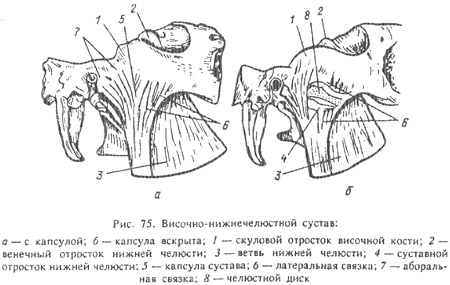
Depending on the type of animal nutrition (herbivores, granivores, carnivores and omnivores), the shape, size of the articular surfaces and the location of the points of attachment of the chewing muscles on the bones, as well as the shape of the rubbing surfaces of the teeth, are very variable. This allows animals to perform grinding, crushing and tearing types of chewing movements of the lower jaw.
Both jaw joint(right and left) work interconnected. The joint is covered with a capsule - capsula articularis, and has an additional lateral ligament - ligamentum laterale. Between the connecting bones there is an articular disc - discus articularis, which is a biconcave oblong-oval plate made of fibrous cartilage. It is inserted between the articular process of the lower jaw and the articular tubercle of the temporal bone. Movement in the joint is limited and directed along two specific axes.
In herbivores, in addition to the lateral ligament, there is also a posterior ligament made of elastic tissue. It goes from the articular process of the temporal bone to the articular process of the lower jaw.
The concept of organs, apparatus and digestive organs
None of the tissues in the body occurs in isolated form. As already noted, epithelial tissues contain elements nerve tissue– nerve fibers, sensory and motor nerve endings; in musculoskeletal tissues there are not only elements of nervous tissue, but also blood vessels, and cartilage, bone and muscle tissues contain elements of nervous tissue, blood vessels and connective tissue. Moreover, in different parts of the body the combination of tissues is different.
A part of the body consisting of certain tissues and performing a special function is called an organ. For example, the liver produces bile, urine is secreted through the kidneys, the lungs serve as a respiratory organ, and the eye serves as an organ of vision. In each organ, one tissue is leading, main, it reflects main function organ, for example, in glands - epithelial tissue, in muscles - muscle. This tissue makes up the parenchyma of the organ. In addition to the parenchyma, each organ contains:
a) sensory and motor nerves, which either enhance the function of the organ parenchyma, or, on the contrary, stop it;
b) through blood and lymphatic vessels, nutrients and oxygen are delivered to the parenchyma of the organ, without which the life and function of parenchyma cells is impossible;
c) blood and lymphatic vessels have their own special nerves - vascular or sympathetic, also sensory and motor. Both the parenchyma of the organ, and the nerves of the parenchyma, and the vessels of the parenchyma with their nerves are packed into a connective tissue framework, which also has its own nerves and its own vessels.
All organs arise during the development of the organism. This determines their shape, the relative position of the relationship in the work. Bodies performing any one general function, form either an apparatus or an organ system. An organ system is characterized not only by a specific function, but also by the initial structure of individual organs. For example, the muscular system consists of muscles; skeletal system - made up of bones and ligaments; nervous system - from nerve cells etc. The device also performs some general function, but consists of different organs, different in their structure and private functions. For example, the digestive apparatus has a common function, but their individual organs are structured differently, and their functions are varied, although purposeful. The same is true for breathing apparatus, urination, movement (from bone and muscular systems), reproduction. An organism is a complex, unified and integral living system in which everything is in strict mutual dependence, and any change in one part causes a corresponding change in other parts. The body is interconnected with specific living conditions. All functions of the animal body are regulated by the activity of the cerebral cortex. Nervous system carries out the relationship between the body and the external environment. An organism exists and develops in certain, specific environmental conditions. Environmental factors cause the occurrence of certain functions, and functions determine the structure of organs. For example, the structural features of teeth and other digestive organs depend on the type of food, and the nature of the coat changes depending on climatic conditions. I.V. Michurin wrote: “Every organ, every property, every member, all internal and external parts of every organism. conditioned by the external environment of its existence.
Structure and characteristics of the brain and facial parts of the skull of various farm animals
The brain and facial parts of the skull are examined. The border between them passes approximately through the posterior edge of the eye sockets. The medulla of the skull contains the brain and is connected to the atlas. In the facial part of the skull are located nasal cavity and organs of the oral cavity.
The skull consists of 13 paired and 7 unpaired bones. The unpaired bones of the brain section of the skull include: occipital, sphenoid and interparietal bones; to the paired ones - parietal, temporal and frontal. The unpaired bones of the facial part of the skull are: ethmoid, vomer, hyoid and proboscis (in pigs), paired are the upper jaw, incisor, nasal, lacrimal. zygomatic, palatine, pterygoid, lower jaw, nasal concha (upper and lower).
The shape of the skull changes dramatically during ontogenesis. In fetuses and newborns, the skull is more rounded, since its cerebral part is more developed. As teeth grow, the facial part of the skull begins to develop more strongly, especially in herbivores. With age, the shape of the cerebral part of the skull also changes, as masticatory muscles. Side functions are also reflected in the shape of the skull. In pigs, the ability to dig the ground determines the powerful development of the jugular processes and the scales of the occipital bone, as a result of which the skull as a whole takes the shape of a tetrahedral pyramid. In cattle, due to the development of horns that serve for protection, the frontal bone is greatly enlarged, which forms the frontal crest between the base of the horns
This ridge forms the posterior upper edge of the skull. In small cattle and other species of animals there is no frontal crest, and the posterior upper edge of the skull is formed by the occipital crest of the occipital bone.
Powerful development of teeth causes overgrowth upper jaws and the formation of maxillary sinuses (cavities) in them, communicating with the nasal cavity. Sinuses increase bone strength. Such sinuses are also present in some other bones of the skull, for example in the frontal bones, especially in horned cows.
Strong development of masticatory muscles causes:
1) the formation of the zygomatic arches, to which the masticatory muscles and lower jaws are attached;
2) the formation of sometimes more or less deep temporal fossae in the cerebral part of the skull;
H) the formation of zygomatic ridges in horses or facial tuberosities in cattle;
4) closure of the eye sockets
Posteriorly, by combining the frontal bones with the zygomatic arches in herbivores, which increases the strength of the jaw joints;
5) strong development of the branches and angles of the lower jaw.
The skull has many holes and canals for blood vessels that supply the organs of the cranial and nasal cavities, and for the cranial nerves that exit the brain.
The structure and topography of the salivary glands, the composition of saliva and its importance in digestion
In the wall of the mucous membrane of the lips, cheeks, tongue, and velum palatine, the parietal salivary glands are embedded in the form of separate formations or groups. Outside the oral cavity there are large salivary glands:
- paired parotid
– sublingual
– submandibular.
The secretion of the salivary glands, which flows into the oral cavity through the excretory ducts, is called saliva. Functionally, the salivary glands are divided into serous, mucous and mixed. The secretion of the serous glands contains a lot of protein, which is why they are also called proteinaceous. The secretion of the mucous glands contains the mucous substance mucin. Mixed glands secrete a protein-mucus secretion.
The parotid salivary gland is serous (mixed in some areas in carnivores), and is of the alveolar type in structure. In cattle, pigs and dogs it is triangular in shape, in horses it is rectangular. Lies at the base auricle. Its excretory duct opens into the vestibule of the oral cavity: in horses and at the level of the 3rd, in cattle - 3-4th, in pigs - 4-5th upper molar.
The submandibular salivary gland is mixed. In cattle, it is relatively long, extending from the atlas to the submandibular space, the excretory duct opens in the sublingual wart at the bottom of the oral cavity. In pigs it is round, covered by the parotid gland; the excretory duct opens in pigs next to the frenulum of the tongue.
The sublingual salivary gland is double. In cattle, the short duct part lies under the mucous membrane of the floor of the mouth, numerous short excretory ducts open on the side of the body of the tongue; the long-ductal part is located next to the previous one, its long excretory duct opens in the sublingual wart. Functionally, the long-duct part is mixed, the short-duct part is mucous. Horses have only a short-ductal part, the secretion is mixed in nature.
Saliva is a mixed secretion of three salivary glands (parotid, submandibular and sublingual), colorless, slightly cloudy (from the presence of mucin), slightly alkaline (especially in ruminants), odorless. Mucin gives it a peculiar consistency and slipperiness, as a result of which the food soaked in saliva is easily swallowed.
Saliva is a solvent for flavoring substances. Its enzymatic role in animals is small. Only pigs' saliva contains two enzymes that break down carbohydrates (starch): amylase converts starch into dextrins, and the latter into the disaccharide maltose; under the influence of the second enzyme - maltose - maltose is split into two particles of grape sugar.
The composition of saliva varies depending on the type and amount of food. As a rule, dry and roughage produce more saliva than wet food. On average, a horse produces 40 liters of saliva per day, a cattle 50–80 liters, and a pig 15 liters. Salivation increases significantly if the food is moistened with a weak solution of table salt.
The abdominal cavity is divided into regions. Serosa abdominal cavity
The abdominal cavity occupies the space between the diaphragm and the pelvis. Posteriorly, it passes into the pelvic cavity without any border. The upper wall of the abdominal cavity is formed by the lumbar and last thoracic vertebrae and the muscles adjacent to them. The lateral and lower walls are represented by the abdominal muscles, and the lower wall is also represented by the xiphoid cartilage. To better understand the position of the organs lying in the abdominal cavity, it is divided into sections. By two conditional segmental planes, the anterior one passing through the last pair of ribs, and the posterior one passing through the vertebrae, the abdominal cavity is divided into anterior, middle and posterior sections.
The anterior abdominal region extends from the diaphragm to the anterior segmental plane. By a plane drawn along the costal arch, this section is divided, in turn, into two subsections - upper and lower. The lower subsection lies between the named plane and the abdominal wall, almost triangular in shape, and is called the region of the xiphoid cartilage. The upper subsection is divided by the midsagittal plane into two equal areas - the right and left hypochondrium.
The middle abdominal section, with two parallel sagittal planes touching the free ends of the transverse costal processes of the lumbar vertebrae, is divided into three subsections - the middle and two lateral. The lateral subsections are called the right and left iliac regions. The middle subsection, in turn, is divided by a conventional frontal plane drawn at the level of the middle shoulder joint, to the upper - lumbar, or renal, and lower - umbilical region. The posterior abdominal region is also divided into three areas: the middle - pubic (pudendal) and the right to left inguinal. Groin areas are a continuation of the iliac regions, and the flax region is a continuation of the umbilical region.
The peritoneum is a serous membrane covering the walls of the abdominal cavity and the peritoneal organs lying in it, lining the walls, called parietal or parietal, the part covering the organs is visceral or splanchnic. Moving from the walls to the organs and from one organ to another, the peritoneum forms doublings and folds; the latter either suspend the organs or connect one to the other. The duplications of the peritoneum are called mesenteries, omentums and ligaments.
The mesentery is a considerable length of doubling of the peritoneum, which suspends the intestine and connects its sections. The mesentery is called after the section of intestines it supports. The place where the mesentery originates from the vertebrae (thoracic and lumbar) is called the root of the mesentery. The horse has two mesenteric roots - anterior and posterior. Animals of other species have only one root. Ligaments are short doublings of the peritoneum that connect organs to each other. Omentums are duplications of the peritoneum, one of which is located between the liver, duodenum and the lesser curvature of the stomach, and the other in the form of an apron goes from the greater curvature of the stomach and protrudes almost to the pelvic cavity. The first duplication is called the lesser omentum, the second - the greater omentum. All organs of the abdominal cavity touch each other and the walls of the cavity with a serous covering. And since in its normal state it is always smooth and moistened with serous fluid, the organs can easily slide around each other.
The process of digestion in the large intestine
Digestion. At the beginning of the large intestine, digestion occurs under the influence of enzymes from the juices of the small intestine and partly from the intestinal juice of the large intestine, which is characterized by a high mucus content and weak enzymes. Intestinal juice is separated here under the influence of mainly mechanical stimuli, mainly indigestible coarse particles of feed. The colon is home to a huge variety of bacteria. This is facilitated by the slower movement of contents through the large intestine compared to the small intestine due to weak peristalsis. Food masses can linger here for up to three days. Among the bacteria of the large intestine, species occupy a special place. fermenting carbohydrates and causing rotting of proteins. As a result of fermentation and decay, organic acids, gases (carbon dioxide, methane, hydrogen sulfide) and toxic substances such as phenol, skatole, cresol, indole are formed. Of the enzymes secreted by bacteria, cellulose breaks down fiber into the disaccharide cellubiose, and the latter, under the influence of cellubiose, breaks down into two molecules of grape sugar. These processes are especially intense in the horse's large intestine. In ruminants, as already mentioned, a significant part of the fiber is broken down in the rumen. Due to the noted structural and functional features digestive tract, ruminants and horses, the digestion of fiber in animals of these species can reach 35–40%, and chemically pure cellulose – 80–90%, while in omnivores it is 15–20%, and in dogs fiber is not digested at all. Microorganisms in the large intestine (and in ruminants, in the rumen) synthesize vitamins B and C. Throughout the large intestine, iron, calcium and magnesium salts are released from the blood through its mucous membrane.
Suction. In the section of the large intestine, water and mineral salts are absorbed in the form of solutions, and in herbivores, nutrients are also absorbed, since a significant amount of food is digested here. Absorption is possible even in the terminal sections of the large intestine.
Formation of feces. The remaining contents of the large intestine after absorption are glued together by mucus secreted by goblet cells of the mucous membrane and formed into feces.
The composition of feces includes:
1) indigestible feed components;
2) substances that are digestible, but have not had time to undergo decomposition;
3) breakdown products of nutrients that have not had time to be absorbed;
4) microbial bodies;
5) remains of digestive juices and waste products of the epithelium of the gastrointestinal tract;
6) excreta of the liver and gastrointestinal canal.
The amount and composition of feces is strongly influenced by the composition of the diet: the more easily digestible substances in the diet, the less feces are formed, and there are few components of the food taken in it.
Defecation (excretion of feces) occurs periodically and is caused by irritation of the sensory nerves of the mucous membrane of the lower colon and rectum by accumulations in them feces. Excitation along the centripetal nerve is transmitted to the center of defecation, located in the lumbosacral part spinal cord, and from there along the centrifugal nerves - to the muscles of the intestines and sphincter anus. A peristaltic wave of the posterior intestine occurs, the internal and external sphincters of the rectum relax, and feces come out.
List of sources used
1. Anatomy and physiology of farm animals. Ed. 4th revision and additional M. "Spike" 1978
2. Anatomy and physiology of farm animals Series: Textbooks and teaching aids for students of secondary specialized educational institutions 2007
Structure and characteristics of the brain and facial parts of the skull of various farm animals
The brain and facial parts of the skull are examined. The border between them passes approximately through the posterior edge of the eye sockets. The medulla of the skull contains the brain and is connected to the atlas. The facial part of the skull contains the nasal cavity and oral organs.
The skull consists of 13 paired and 7 unpaired bones. The unpaired bones of the brain section of the skull include: occipital, sphenoid and interparietal bones; to the paired ones - parietal, temporal and frontal. The unpaired bones of the facial part of the skull are: ethmoid, vomer, hyoid and proboscis (in pigs), paired are the upper jaw, incisor, nasal, lacrimal. zygomatic, palatine, pterygoid, lower jaw, nasal concha (upper and lower).
The shape of the skull changes dramatically during ontogenesis. In fetuses and newborns, the skull is more rounded, since its cerebral part is more developed. As teeth grow, the facial part of the skull begins to develop more strongly, especially in herbivores. With age, the shape of the cerebral part of the skull also changes, as the masticatory muscles are attached to it. Side functions are also reflected in the shape of the skull. In pigs, the ability to dig the ground determines the powerful development of the jugular processes and the scales of the occipital bone, as a result of which the skull as a whole takes the shape of a tetrahedral pyramid. In cattle, due to the development of horns that serve for protection, the frontal bone is greatly enlarged, which forms the frontal crest between the base of the horns
This ridge forms the posterior upper edge of the skull. In small cattle and other species of animals there is no frontal crest, and the posterior upper edge of the skull is formed by the occipital crest of the occipital bone.
The powerful development of teeth causes the growth of the upper jaws and the formation of maxillary sinuses (cavities) in them, communicating with the nasal cavity. Sinuses increase bone strength. Such sinuses are also present in some other bones of the skull, for example in the frontal bones, especially in horned cows.
Strong development of masticatory muscles causes:
1) the formation of the zygomatic arches, to which the masticatory muscles and lower jaws are attached;
2) the formation of sometimes more or less deep temporal fossae in the cerebral part of the skull;
H) the formation of zygomatic ridges in horses or facial tuberosities in cattle;
4) closure of the eye sockets
Posteriorly, by combining the frontal bones with the zygomatic arches in herbivores, which increases the strength of the jaw joints;
5) strong development of the branches and angles of the lower jaw.
The skull has many holes and canals for blood vessels that supply the organs of the cranial and nasal cavities, and for the cranial nerves that exit the brain.