Sphenoid bone, os sphenoidale, unpaired, resembles a flying insect, which explains the name of its parts (wings, pterygoid processes).
The sphenoid bone is the product of the fusion of several bones that independently exist in animals, therefore it develops as a mixed bone from several paired and unpaired ossification points, forming 3 parts at the time of birth, which in turn fuse into a single bone by the end of the first year of life.
It has the following parts:
1) body, corpus;
2) big arms, alae majores;
3) small wings,alae minores;
4)pterygoid processes, processus pterygoidei(its medial plate is the former double pterygoid, develops on the basis of connective tissue, while all other parts of the bone arise on the basis of cartilage).
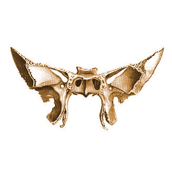
Sphenoid bone. Rear view. 1. Visual channel; 2. Saddle back; 3. Posterior inclined process; 4. Anterior inclined process; 5. Small wing; 6. Superior orbital fissure; 7. Parietal angle; 8. Large wing (cerebral surface); 9. Round hole; 10. Pterygoid canal; 11. Scaphoid fossa; 12. Lateral plate (pterygoid process); 13. Pterygoid notch; 14. Groove of the pterygoid hook; 15. Vaginal process; 16. Wedge-shaped ridge; 17. Body of the sphenoid bone; 18. Medial plate (pterygoid process); 19. Wing-shaped hook; 20. Pterygoid fossa; 21. Internal groove carotid artery.
Body, corpus, on its upper surface has a depression along the midline - saddle turcica, sella turcica, at the bottom of which lies hole For pituitary gland, fossa hypophysialis.In front of it there is an elevation, tuberculum sellae, along which passes transversely sulcus chiasmatis for cross ( chiasma) optic nerves; at the ends sulcus chiasmatis visible visual channels, canales optici, through which the optic nerves pass from the cavity of the orbits to the cavity of the skull. Posteriorly, the sella turcica is limited by a bony plate, back of the saddle, dorsum sellae. On the lateral surface of the body there is a curved carotid sulcus, sulcus caroticus, trace of the internal carotid artery.
On the anterior surface of the body, which is part of the posterior wall of the nasal cavity, is visible crest, crista sphenoidalis, below entering between the wings of the opener. Christa sphenoidalis connects anteriorly with the perpendicular plate of the ethmoid bone. Irregularly shaped holes are visible on the sides of the ridge, aperturae sinus sphenoidalis, leading to air sinus, sinus sphenoidalis, which is placed in the body of the sphenoid bone and is divided partition, septum sinum sphenoidalium, in two halves. Through these openings the sinus communicates with the nasal cavity. In a newborn, the sinus is of very small size and only around the 7th year of life begins to grow rapidly.
Small wings, alae minores, are two flat triangular plates, which with two roots extend anteriorly laterally from the anterosuperior edge of the body of the sphenoid bone; between the roots of the small wings are the mentioned visual channels, canales optici. Between the lesser and greater wings there is the superior orbital fissure, fissura orbitalis superior, leading from the cranial cavity to the orbital cavity.
Big wings, alae majores, extend from the lateral surfaces of the body laterally and upward. Near the body, behind fissura orbitalis superior available round hole, foramen rotundum, leading anteriorly into the pterygopalatine fossa, caused by the passage of the second branch trigeminal nerve, n. trigemini. At the back, a large wing in the form of an acute angle protrudes between the scales and the pyramid of the temporal bone. There is a foramen spinosum, foramen spinosum, through which it passes a. meningea media. Much more is visible in front of him foramen ovale, foramen ovale, through which the third branch passes n.trigemini.
Large wings have four surfaces: brain,facies cerebralis, orbital,facies orbitalis, temporal, facies temporalis, And maxillary, facies maxillaris. The names of the surfaces indicate the areas of the skull where they face. The temporal surface is divided into the temporal and pterygoid parts by infratemporal crest, crista infritemporalis.
Pterygoid processes, processus pterygoidei extend vertically downward from the junction of the greater wings and the body of the sphenoid bone. Their base is pierced by a sagittal canal, canalis pterygoideus, - the place of passage of the named nerve and vessels. The anterior opening of the canal opens into the pterygopalatine fossa.
Each process consists of two plates - lamina medialis And lamina lateralis, between which a fossa, fossa pterygoidea, is formed at the back.
The medial plate is bent at the bottom crochet, hamulus pterygoideus, through which the tendon that begins on this plate is thrown m. tensor veli palatini(one of the muscles of the soft palate).
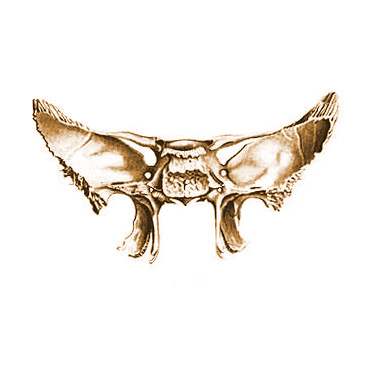
Sphenoid bone. Front view. 1. Aperture of the sphenoid sinus; 2. Saddle back; 3. Wedge-shaped shell; 4. Small wing; 5. Superior orbital fissure; 6. Zygomatic edge; 7. Infratemporal surface, 8. Spine of the sphenoid bone; 9. Pterygopalatine groove; 10. Lateral plate; 11. Wing-shaped hook; 12. Medial plate of the pterygoid process; 13. Vaginal process; 14. Wedge-shaped ridge; 15. Pterygoid notch; 16. Pterygoid canal; 17. Round hole; 18. Infratemporal crest; 19. Orbital surface big wing; 20. Temporal surface of the greater wing.
Variants and anomalies
Failure of fusion of the anterior and posterior halves of the body of the sphenoid bone leads to the formation of a narrow, so-called craniopharyngeal canal, in the center of the sella turcica. The foramen ovale and foramen spinosum sometimes merge into one common foramen; the foramen spinosum may be absent.
At the center of the base of the skull lies the sphenoid bone. The pits and cavities of the facial and cerebral parts of the skull, as well as the lateral walls of the cranial vault, are the anatomical formations in which it participates.
In terms of its structure, the sphenoid bone of the skull is quite complex. It is formed by a body from which three paired processes extend in different directions:
- Pterygoid processes;
- Small wings;
- Big wings.
Body of the sphenoid bone
Geometrically, the body of the sphenoid bone of the skull resembles the shape of a cube with irregular sides. The sphenoid sinus is the name given to the cavity that is located inside this cube.
The body of the sphenoid bone has 6 surfaces:
- Anterior, passing into the lower surface without boundaries and restrictions
- A pair of side surfaces
- Posterior (which in an adult is fused with the main surface of the occipital bone)
- Upper (cerebral)
A distinctive feature of the upper, or so-called “cerebral” surface of this bone, is the presence of a special anatomical formation, called the “sella turcica”. It is mainly important and “famous” for the fact that inside this formation there is a pituitary fossa. As the name implies, this is where a special hormonal gland is located - the pituitary gland. Anterior to this depression is the tubercle sella, located transversely. Visually, that part of the sella turcica, which is called the back, stands out quite strongly. Its lateral parts protrude forward, forming, as it were, a pair of inclined processes. The base of the back of the sella turcica is characterized by the presence of another special anatomical formation.
 We are talking about the so-called “carotid sulcus” - this is a special opening through which one of the most important arteries of our body, the carotid artery, passes. A little posteriorly there is a so-called “wedge-shaped tongue”, whose structure turns the carotid sulcus into a kind of deep groove. Together with the apical part of the pyramid of the temporal bone, this formation is a kind of internal limiter of the carotid foramen. Through this hole, the internal carotid artery opens into the cranial cavity from the carotid canal.
The anterior surface of the body of the sphenoid bone is elongated and forms a wedge-shaped ridge. Characteristic feature The wedge is the formation on its sides of special bone plates - wedge-shaped shells, which limit the openings of the sphenoid sinus.
From the anterior surface of the body of the sphenoid bone of the skull, a large wing begins, which is a pair. The trio of holes on each wing lies at its very base. Through them the middle meningeal artery and branches penetrate into the cranial cavity trigeminal nerve(2nd and 3rd).
Greater wing of the pterygoid
In the large wing of the pterygoid bone of the skull, it is customary to distinguish four surfaces:
- Brain. Differs in the severity of arterial grooves and finger-like impressions;
- Maxillary. It is located in the form of a triangular section, starting at the top of the orbital surface and the base of this “triangle” ending at the bottom with the pterygoid process. A round hole opens on the surface;
- Temporal. This is the largest surface of the large wing of the pterygoid bone of the skull, divided into two parts by the infratemporal crest. The temporal fossa includes the largest, upper part of this surface, which is located almost vertically. The horizontally located small part of the surface participates in the formation of the small wall of the infratemporal fossa;
- Orbital. This surface is part of the lateral wall of the orbit and has a quadrangular shape.
Lesser wing of the pterygoid
When talking about the small wing of the sphenoid bone of the skull, one cannot fail to mention its structure. These are paired plates located on the sides of the body of this bone. Between their roots is the optic canal. Along it, the optic nerve emerges from the orbit. On the anterior edges of the small wings there are serrations for connection with the orbital part of the frontal bone and the plate of the ethmoid bone. The rear edges of the wings do not have such notches.
Small wings of yours top surface participate in the formation of the cranial cavity, their lower part participates in the formation of the orbit. The superior orbital fissure is the special space between the greater and lesser wings. Through this anatomical formation, the abducens, oculomotor, and lateral nerves, as well as the optic nerves, enter the orbit.
Wedge-shaped paired process
A paired pterygoid process extends vertically down from the origin of the large wing of the pterygoid bone of the skull. With its medial plate it faces the nasal cavity, and with its lateral plate it faces the infratemporal fossa.
The structure of the base of this process is characteristic. It has a very narrow canal through which nerves and blood vessels pass. We are talking about the pterygoid canal. On the pterygoid process of the pterygoid bone of the skull, it is customary to distinguish medial and lateral plates. They are fused at the front. They diverge posteriorly, forming a pterygoid fossa. Below they are separated by the so-called pterygoid notch. The medial plate of this process below forms a pterygoid hook and is narrower and longer than the lateral plate.
17. Sphenoid bone, its parts, holes, channels and their contents.
sphenoid bone,os sphenoidale, located in the center of the base of the skull. It participates in the formation of the lateral walls of the cranial vault, as well as the cavities and fossae of the brain and facial departments skulls The sphenoid bone has a complex shape and consists of a body from which 3 pairs of processes extend: large wings, small wings and pterygoid processes.
Body,corpus, The sphenoid bone has the shape of an irregular cube. Inside it there is a cavity - the sphenoid sinus, sinus sphenoidalis. There are 6 surfaces in the body: the upper, or cerebral; posterior, fused in adults with the basilar (main) part of the occipital bone; the front one, which passes without sharp boundaries into the lower one, and two lateral ones.
Small wing, ala minor, It is a paired plate extending from each side of the body of the sphenoid bone with two roots. Between the latter is the visual channel, canalis opticus, for the passage of the optic nerve from the orbit. The anterior edges of the lesser wings are serrated; the orbital parts of the frontal bone and the cribriform plate of the ethmoid bone are connected to them. The posterior edges of the small wings are free and smooth. On the medial side of each wing there is an anterior inclined process, processus clinoideus anterior. The dura mater of the brain grows to the anterior as well as to the posterior inclined processes.
The lesser wing has an upper surface facing the cranial cavity, and a lower one, participating in the formation of the upper wall of the orbit. The space between the lesser and greater wings is the superior orbital fissure, fissura orbitalis superior. The oculomotor, lateral and abducens nerves (III, IV, VI pairs) pass through it from the cranial cavity to the orbit cranial nerves) And optic nerve- I branch of the trigeminal nerve (V pair).
Big wing, ala major, paired, begins with a wide base from the lateral surface of the body of the sphenoid bone (Fig. 32). At the very base, each wing has three holes. Above the others and in front there is a round hole, foramen rotundum, through which the second branch of the trigeminal nerve passes, in the middle of the wing there is the foramen ovale, foramen ovale, for the third branch of the trigeminal nerve. Foramen spinosum, foramen spinosum, smaller in size, located in the region of the posterior corner of the large wing. Through this opening, the middle meningeal artery enters the cranial cavity.
The large wing has four surfaces: medullary, orbital, maxillary and temporal. On the surface of the brain fades cerebralis, finger-shaped impressions are well defined, impressidnes digitatae, and arterial grooves, sulci arteriosi. orbital surface, fades orbitalis, - quadrangular smooth plate; part of the lateral wall of the orbit. maxillary surface, fades maxillaris, occupies a triangular area between the orbital surface above and the base of the pterygoid process below. On this surface, facing the pterygopalatine fossa, a round opening opens. Temporal surface, fades tempordlis, the most extensive. infratemporal crest, crista infratempo- ralis, divides it into two parts. Upper part larger in size, located almost vertically, part of the wall of the temporal fossa. The lower part is located almost horizontally and forms the upper wall of the infratemporal fossa.
pterygoid process,processus pterygoideus, paired, departs from the body of the sphenoid bone at the beginning of the large wing and is directed vertically downward. The medial plate of the process faces the nasal cavity, the lateral plate faces the infratemporal fossa. The base of the process is pierced from front to back by a narrow pterygoid canal, canalis pterygoideus, in which blood vessels and nerves pass. The anterior opening of this canal opens into the pterygopalatine fossa, the posterior one - on the outer base of the skull near the spine of the sphenoid bone, splna ossis sphenoidalis. The plates of the pterygoid process are distinguished: medial, lamina medlis, and lateral, lamina lateralis. The anterior plates are fused. Posteriorly, the plates of the pterygoid process diverge, forming the pterygoid fossa, fossa pterygoidea. At the bottom, both plates are separated by a pterygoid notch, incisura pterygoidea. The medial plate of the pterygoid process is somewhat narrower and longer than the lateral one and below passes into the pterygoid hook, hamulus pterygoideus.
- Pterygoid process, processus pterygoideus. Rice. A, B.
- Lateral plate [of the pterygoid process] lamina lateralis. Rice. A, B.
- Medial plate [of the pterygoid process], lamina medialis. Rice. A, B.
- Pterygoid notch, incisura pterygoidea. It is located between the two plates of the pterygoid process and is directed downward. Filled with the pyramidal process os palatinum. Rice. A.
- Pterygoid fossa, fossa pterygoidea. Located between the lateral and medial plastics. Place of attachment of m.pterygoideus medialis. Rice. A, B.
- Scaphoid fossa, fossa scaphoidea. Depression at the base of the medial plate of the pterygoid process. The starting point of mjensor veli palatini. Rice. A.
- Vaginal process, processus vaginalis. Located from inside base of the medial plate of the pterygoid process. Rice. A, B.
- Palatovaginal groove, sulcus palatovaginal. Together with the palatine bone, it forms the canal of the same name. Rice. B.
- Vomerovaginal groove, sulcus vomerovaginal. It is located at the base of the pterygoid process and, together with the vomer, forms the canal of the same name. Rice. B.
- Wing-shaped hook, hamulus pterygoideus. It is located at the end of the medial plate of the pterygoid process and is directed downward. Rice. A, B.
- Furrow of the pterygoid hook, sulcus hamuli pterygoidei. Formed due to a sharp bend of the wing-shaped hook. Rice. B.
- Pterygoid [[vidian]] canal, canalis pterygoideus []. It passes at the base of the pterygoid process towards the pterygopalatine fossa. Contains the large and deep petrosal nerves Fig. A. See fig. IN.
- Pterygospinous process, processus pterygospinosus. A sharp projection on the posterior edge of the lateral plate of the pterygoid process. Rice. A.
- Temporal bone, os temporale. Located between the occipital, sphenoid and parietal bones. Consists of stony, drum and scaly parts. Rice. B, G, D.
- Pyramid ( rocky part), pars petrosa. Contains the organ of hearing and balance. Rice. G.
- Occipital margin, margo occipitalis. Connects to the occipital bone. Rice. V, G.
- Mastoid process, processus mastoideus. Located behind the external auditory canal. Rice. V, D.
- Mastoid notch, incisura mastoidea. Located on the lower surface of the pyramid, medial to the mastoid process. Place of origin of the posterior abdomen m.digastricus. Rice. IN.
- The groove of the sigmoid sinus, sulcus sinus sigmoidei. Rice. G.
- Groove of the occipital artery, sulcus a.occipitalis. Located at the occipital edge of the pyramid, the medial mastoid notch. Rice. IN.
- Mastoid foramen, foramen mastoideum. Located behind the mastoid process. Contains an emissary vein. Rice. V, G.
- Facial canal, canalis facialis. It begins in the internal auditory canal and ends with the stylomastoid foramen. Contains the nerve of the same name. Rice. B, G, D.
- The genu of the facial canal, geniculum canalis facialis. The bend of the facial canal at the anterior wall of the pyramid, near the cleft of the greater petrosal nerve. Rice. G.
- The canaliculus chordae tympani. A narrow passage connecting the facial canal and tympanic cavity. Contains drum string. Rice. G, D.
- The tip of the pyramid, apex partis petrosae. Directed forward and medially. Rice. V, G.
- Sleepy canal, canalis caroticus. It begins on the outer base of the skull between the jugular foramen and the myotubal canal. Contains the internal carotid artery. Rice. IN.
- Carotid-tympanic tubules, canaliculi caroticotympanic. They pass through the wall of the carotid canal. Contains vessels and nerves going into the tympanic cavity. Rice. IN.
- Musculo-tubal canal, canalis musculotubarius. It is located in front of the carotid canal and leads into the tympanic cavity. Contains the auditory tube and the tensor muscle eardrum. Rice. V, D.
- The semicanal of the tensor tympani muscle, semicanalis m.tensoris tympani. Rice. D.
- Half-channel auditory tube, semicanalis tubae auditoriae (auditivae). Rice. D.
- The septum of the muscular-tubal canal, septum canalis musculotubarii. Bone wall, between the above-mentioned hemicanals. Rice. D.
- Sphenoid bone, os sphenoidale. Located between the frontal, occipital and temporal bones. Rice. A, B, C.
- Body, corpus. Located between the large wings. Rice. A, B.
- Wedge-shaped eminence, jugum sphenoidale. Connects the lesser wings of the sphenoid bone. Rice. A.
- (Pre)cross groove, sulcus prechiasmaticus. Located between the right and left visual channels. Rice. A.
- Turkish saddle, sella turcica. A fossa located above the sphenoid sinus. Contains the pituitary gland. Rice. A.
- Tubercle sellae, tuberculum sellae. Elevation anterior to the pituitary fossa. Rice. A.
- [Middle inclined process, processus clinoideus medius]. Located on the side of the pituitary fossa. Not constantly present. Rice. A.
- Pituitary fossa, fossa hypophysialis. Filled with the pituitary gland. Rice. A.
- The back of the saddle, dorsum sellae. Located posterior to the pituitary fossa. Rice. A, V.
- Posterior inclined process, processus clinoideus posterior. Bilaterally located projections of the back of the saddle. Rice. A, V.
- Carotid groove, sulcus caroticus. It starts from the middle of the torn hole and goes forward. The internal carotid artery passes through it. Rice. A.
- Wedge-shaped tongue, lingula sphenoidalis. Located lateral to the entry point of the internal carotid artery into the skull. Rice. A.
- Wedge-shaped crest, crista sphenoidalis. Located in the midline on the anterior surface of the body and serves as the attachment point for the perpendicular plate of the ethmoid bone. Rice. IN.
- Wedge-shaped beak, rostrum sphenoidale. It is a continuation of the wedge-shaped ridge downwards. Connects to the opener. Rice. IN.
- Sphenoid sinus, sinus sphenoidalis. Paired air cavity of the skull. Rice. IN.
- The septum of the sphenoid sinuses, septum intersinuale sphenoidale. Separates the right sphenoid sinus from the left. Rice. IN.
- Aperture of the sphenoid sinus, apertura sinus sphenoidalis. Opens into a wedge-ethmoid recess. Rice. IN.
- Wedge-shaped shell, concha sphenoidalis. Usually a paired concave plate fused with the body of the sphenoid bone. Forms the anterior and lower walls of her sinus. Rice. IN.
- Small wing, ala minor. Rice. A B.V.
- Optic canal, canalis opticus. Contains the optic nerve and ophthalmic artery. Rice. A.
- Anterior inclined process, processus clinoideus anterior. Paired conical projection of the lesser wings in front of the pituitary fossa. Rice. A.
- Superior orbital fissure, fissura orbitals superior. Located between the large and small wings. Nerves and veins pass through it. Rice. A, B, C.
- Large wing, ala major. Rice. A, B, C.
- Brain surface, fades cerebralis. Facing the brain. Rice. A.
- Temporal surface, fades temporalis. Facing outwards. Rice. B, V.
- Maxillary surface, fades maxillaris. Pointed to the side upper jaw. There is a round hole on it. Rice. IN.
- Orbital surface, fades orbitalis. Facing inside the eye socket. Rice. IN.
- Zygomatic margin, margo zygomaticus. Connects to the zygomatic bone. Rice. IN.
- Frontal edge, margo frontalis. Articulates with the frontal bone. Rice. A.
- Parietal edge, margo parietalis. Connects to the parietal bone. Rice. IN.
- Scaly margin, margo squamosus. The scaly suture articulates with temporal bone. Rice. A.
- Infratemporal crest, crista infratemporalis. It is located between the vertically oriented temporal and horizontally oriented inferior surfaces of the greater wing. Rice. B, V.
- Round hole, foramen rotundum. Opens into the pterygopalatine fossa. Contains the maxillary nerve. Rice. A, B, C.
- Oval hole, foramen ovale. Located medially and anterior to the foramen spinosum. The mandibular nerve passes through it. Rice. A, B.
- [Venous opening, foramen venosum]. Located medial to the foramen ovale. Contains an emissary vein originating from the cavernous sinus. Rice. A, B.
- Spinous foramen, foramen spinosum. Located lateral and posterior to the foramen ovale. Designed for the middle meningeal artery. Rice. A, B.
- [Rocky hole, foramen petrosum, []. Located between the foramen ovale and the foramen spinosum. Contains n.petrosus major. Rice. A, B.
- Spine of the sphenoid bone, spina ossis sphenoidalis. Departs from the large wing and is directed downward. Rice. A, B.
- The groove of the auditory tube, sulcus tubae auditoriae (auditivae). Located on the lower surface of the greater wing, lateral to the base of the pterygoid process. Contains the cartilaginous part of the auditory tube. Rice. B.