The clavicle is the only human bone that holds the skeleton of the torso and upper limb together. It comes from the same elements as the cranial bones. In terms of shape it should be classified as tubular bones, and in structure as spongy bones. It is covered on top by a shell of compact bone. It receives in the sixth week of fetal development in the womb, that is, before all other bones.
Joint stability is regulated by the fibroarthilatory disc and accessory ligaments, which limit both medial displacement and height of the medial end of the clavicle. At the acromioclavicular joint, the lateral end of the clavicle is hinged to the deep facet of the acromion. The articular capsule is inserted into the edges of the articular surfaces. Joint stability is primarily regulated by the powerful coracoclavicular ligament, which links the coracoid process to the inferior aspect of the clavicle.
The lateral end of the clavicle can rise or fall and move forward and backward. The axes of these movements are located in the articular and coracoclavicular ligaments, and not only through clavicular joints. Thus, the medial end of the clavicle rises during the descent of the scapula and moves posteriorly as the scapula moves forward. Complete redness of the upper extremity requires rotation of the scapula so that the glenoid cavity tilts upward. The 40° rotation of the clavicle at the tendon joint complements the 20° of motion available at the acromioclavicular joint and allows the scapula to rotate through an arc of approximately 60°.
Anatomy
The clavicle is an S-shaped bone with a body and two ends:
- the sternal end faces the manubrium of the sternum;
- the acromial end connects to the acromin.
The sternal end, as well as the part of the body adjacent to it, is curved with a convexity forward, the other part is curved with a convexity backward. The middle section of the bone, which is located between its ends, is slightly compressed from top to bottom. Its lower surface has a nutrient opening. At the sternal end there is a depression of the costoclavicular ligament. The humeral end is equipped with a cone-shaped tubercle and a trapezoidal line. Towards the acromial end of the lower surface of the bone there is a groove for the subclavian muscle.
Shoulder joint The shoulder joint is a synovial type of enarrosis and provides wide range movements. Hemispherical head humerus oriented inward and backward and articulates with the pharyngeal cavity of the scapula, which is much smaller in size. The synovial membrane surrounds the fibrous capsule and covers the intracapsular part of the humeral shaft. The movements are: flexion, extension, abduction, adduction, internal rotation and external rotation. Ulnar ulnar ulnar synovial joint between the distal end of the humerus and the proximal ends of the radius and ulna.
The top surface is smooth. The sternal end is thicker. On the inner surface it bears the sternal articular surface. The acromial end is wider than the sternal end, however, it is not so thick. Outside, on its lower part there is an acromial articular surface, which articulates with the acrominus of the scapula.
The anatomy of the clavicle allows us to see the connection of this bone with the human skeleton and its parts.
Surface shoulder joint formed with a slightly concave surface of the radius head. Inward shoulder brachialis muscle tapers into the deep greater sigmoid cavity of the ulna. The capsule is enveloped in a synovial membrane, as well as fatty projections of the radial, coronoid and olecranon pits. There are two collateral ligaments, external and internal. The determining factor for joint stability is the integrity of these ligaments, which firmly hold the connection between the humeral troche and the greater sigmoid cavity.
IN elbow joint Only flexion and extension movements occur. Radial-ulnar joints In the proximal and distal radial joints, synovial, radius and ulnar joints. At the proximal joint, the head of the radius is articulated with the sigmoid cavity of the ulna. The distal radiolumaric junction occurs between the head of the ulna and the sigmoid cavity of the radius. At the level of these joints, the movements of supination and pronation can describe an arc of about 180°. During pronation, the radius rotates across the elbow and rotates the forearm and arm so that the palm faces backward.
For example, the connection between the clavicle and the scapula occurs through the humeral end of the bone and the humeral process of the scapula, which together make up the acromioclavicular joint. Its articular surfaces are beveled, flat and elliptical in shape. This joint is surrounded by a dense fibrous capsule, which is supported by strong ligaments. The sternoclavicular joint is surrounded by a wide fibrous capsule and three powerful ligaments. In this joint, movements can be carried out along three axes perpendicular to each other. Its area is not covered by muscles. For active movements in it, the muscles that are attached to the collarbone are included in the work. The sternal end of the clavicle has two types of muscles:
Supination places the limb segment back into its anatomical position. The axis of movement passes through the head of the radius and the styloid process of the ulna. Articulation of the wrist Mobility of the hand on the forearm is possible thanks to a group of synovial joints. Almost all movement occurs at the radiocarpal joint, to which they complement the movements between the carpal bones themselves. Radiocarpal compound. In this joint, the distal end of the radius with its articular disc is articulated with the bones of the proximal series of carp, which includes from outside to inside, scaphoid, lunate and pyramidal.
- sternocleidomastoid muscle;
- The sternoclavicular part of the pectoral muscle.
The acromial end also moves due to two muscles:
- deltoid muscle;
- trapezius muscle.
The posterior-inferior surface of the bone is equipped with the subclavian muscle. If the joints of the bone and the muscles that are adjacent to it do not move, it will be impossible to carry out active movements of the arms.
The capsule is inserted into the edges of the articular surfaces and strengthened by lateral connections. Carp joints. The bones of the carp are arranged in two rows: the proximal row, formed from the outside to the inside by the cascade, lunate, pyramidal and chest, and the distal row, consisting of the trapezium, trapezium, big bone and inhabitant The joints between the carpal bones are strengthened by anterior, posterior and interaxial connections. The cavities of these joints are usually interconnected and function as a single unit, which is called “mediocarpal articulation”.
The movements of the radiocarpal and intercarpal joints are complementary and allow flexion, extension, adduction and abduction of the forearm. The carpometarcar, metacarpophalangeal, and interphalangeal joints of the hand are synovial and allow for a variety of hand functions, including complex movements associated with grasping objects. Carpometacarpal joints There are three independent carpometacarpal joints, one for the thumb and two for the other fingers. The articulation between the first metacarpal and the trapezius has articular surfaces of the joint.
Functions
There are three main functions of the clavicle.
- Support function. As we have already said, there is a direct connection between the clavicle and the scapula, which is confirmed by the anatomy of the bone. It is on it that the scapula, as well as the upper limb, are suspended. In addition, the bone connects the arm and body, thereby providing maximum mobility.
- Participates in hand-to-hand transmission axial skeleton physical impulses.
- Protects lymphatics, blood vessels and the nerves that are located between the neck and arm.
As you can see, this bone carries out important role in the body. However, it is susceptible to various injuries that affect human activities.
This articulation allows movements of flexion and extension in a plane parallel to the palm, as well as adduction and abduction in a plane perpendicular to the plane. During the opposition thumb rotates so that he can touch it with any fingertip. The other two carpometarcal joints are flat and less mobile than the first. Metacarpophalangeal joints These joints occur between the rounded heads of the metacarpals and the concave bases of the proximal phalanges. Each of the connections is strengthened by lateral connections.
The joints allow movements of flexion, extension, adduction and abduction. Because the collateral ligaments are tense during flexion, adduction and abduction movements can only be performed in a position of extension. Specific architecture lower limb allows us, unlike other mammals, to support the weight of our body on two limbs, at the same time they serve for locomotion, and at all times the body weight is distributed, adapting to the situation. The entire lower limb is responsible for determining the foot in space.
Damage
Three groups of clavicle injuries can be distinguished.
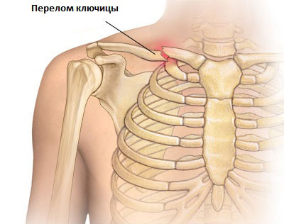
- Fracture. Most often, a fracture occurs in the area of the diaphysis, that is, the middle of the bone body. Since there is a left and a right collarbone, the damage is localized to one of them, or in rare cases to both bones. Sometimes damage to the collarbone and scapula, joints occurs, and rupture of the muscles and ligaments that surround the bone occurs. From the anatomy of the bone, it is clear that much is interconnected in this area, so damage to several parts often occurs at once. The causes of fractures are most often direct trauma, which occurs due to a fall on the arm or as a result of direct blow. A bone fracture can occur in newborns and is quite common. This happens as the fetus passes through the birth canal. Signs of a fracture include lengthening of the limb, crepitus and deformity, and inability to raise the arm.
- acromioclavicular joint. The cause of this injury is a fall on the shoulder. At this point, the scapula comes off the bone and dislocation occurs. In this case, there is a strong elongation of the upper limb, swelling and deformation, as well as the “key” symptom. The last sign is characterized by the end of the clavicle and its return to a state of pathology.
- bones. This rare pathology, which is associated with resorption bone tissue. The causes of this disease are not clear, but they are associated with autoimmunization of bone tissue. At the same time, the collarbone does not hurt. Clinically, the disease manifests itself as poorly healing fractures. Again, this can affect either the right or left side.
Treatment and prevention
It is very important to provide first aid to the injured person. You need to hang your hand on a scarf. You can bandage it to the body, while the elbow joint should be bent at a right angle. The patient should then be taken to the hospital or an ambulance called for him.
The lower limb consists of four segments: the pelvic girdle, the thigh, the leg and the leg. The pelvic girdle is formed by both the coxal bones and the sacrum. It is a structure of great solidity, which contains the internal genital organs and the terminal parts of the urinary and digestive apparatus. It serves as an introduction to the muscles responsible for upright posture, the abdominal wall, and the lower limb. The hip has only one bone, the femur, which is connected to the pelvic girdle by the hip joint and the leg by the knee joint.
The leg is formed by two bones, the inner, tibia, and the outer, fibula, which are articulated by the upper and lower tibiofibular joints. Hip joint It is a spherical synovial joint between the head of the femur and the cavity acetabulum hips. The head of the femur is lined with hyaline cartilage. The head protrudes from the neck of the femur, the base of which ends at the face of the greater trochanter. In the deep cotiloid cavity, the articular surfaces are covered with hyaline cartilage, and the round ligaments are inserted at the edges.
Treatment of a fracture depends on the nature and severity. Subosseous and non-displaced fractures are treated with fixation bandages. They are applied for several weeks in children; in adults, the period increases to one month. If the fragments are displaced, local anesthesia is first performed, then the fragments are reduced. It is quite difficult to keep them in the desired position, so methods for fixing them have been developed. For example, there are Delbe rings that grip the shoulder joints. They connect at the back.
The idiofemoral, frontal, and ischiofemoral ligaments are capsular thickenings that spiral downwards from the coxal bone to the femur. Its base is inserted into the iliac spine and its two shoulders, at the ends of the interline. The ischiofemoral is located on the posterior side and reaches the base of the greater trochanter. Inside the joint is a round ligament with a flattened cone shape, the base of which is inserted into the edge of the acetabular cavity.
Its apex is at the head of the femur. The synovial membrane covers the inside of the capsule and the incompatible surfaces of the joint, it also covers the round ligament and is reflected on the reticular fibers and the neck of the femur towards the head. Movements. The hip joint is multi-axial and allows movements of flexion, extension, adduction, abduction, external and internal rotation and circumambulation. Stability This articulation owes much to its robust stability to the morphology of the bones that compose it and to the deep situation of the femoral head in the acetabulum cavity.
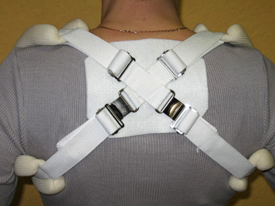
If the collarbone is injured in such a way that prolonged bed rest is required, the patient should lie on a bed with a firm mattress and the arm should be hung down and behind. If damaged soft fabrics, are carried out surgical operations, which help restore bone function. The operation, which is accompanied by the combination of fragments, is called osteosynthesis. It is based on bone restoration. During the process, fixation is performed with a special plate. For fractures, a wire may be installed to hold the bone in the correct position.
Other factors that also influence are the ligaments and the tone of the muscles that cross the joint. Knee joint The knee joint is a synovial joint between the distal end of the femur, the proximal end of the tibia, and the posterior aspect of the patella. It is a loose joint with a wide degree of flexion and extension and limited internal and external rotation. It is a relatively superficial joint on its anterior and lateral sides, where some of the bony structures can be palpated.
On the other hand, its posterior part, located deep in the popliteal cavity, cannot be palpated. On the anterior side, the patellar ligament extends between the apex of the patella and the anterior tuberosity of the tibia. It can be easily palpated and used clinically to examine the stretch reflex. The connecting surfaces are the outer and inner protrusions of the femur, which are covered with hyaline cartilage. These surfaces taper to the corresponding tibial condyles.
Primary prevention is concerned with preventing damage. You need to be careful while walking, especially on slippery surfaces. You should also strengthen your bones. Secondary prevention aims to prevent late complications, which include malunion of the clavicle and subsequent deformation shoulder girdle. To avoid this, you need to contact a traumatologist as soon as possible. Timely treatment and high-quality rehabilitation will help you quickly return to your previous rhythm of life.
The vascular surface for the patella is located in the anterior aspect of the femur. Posteriorly, a deep intercondylic notch separates the condyles. The articular capsule, thin on its anterior and posterior sides, has strong reinforcement of the collateral ligaments on its sides. Toward the sides of the femur, the capsular insertion extends to the epicondyles. Posteriorly, it is inserted into the upper edge of the condyles and into the intercondiral line. On the sides of the tibia, a capsule is inserted near the edges of the joints. The lateral portions of the capsule that loosely attach to the superficial edges of the meniscus at the femur and tibia are called coronary ligaments.
There is no official diagnosis for scapular dislocation. There are 2 dislocations in this part of the body: in the shoulder and scapula. Doctors explain this by the fact that in any dislocated place there is a peripheral bone, and in our case it will be the humerus. The exception to the rule is the collarbone. There are dislocations of the sternal or scapular end of the clavicle, but not of the scapula.
Meniscus The inner and outer meniscus are crescent shaped. Its front and rear horns are inserted into the intercondiral surface of the tibia, and its superficial edges are attached to the joint capsule. They vary in size and shape, the inner one being the narrowest and somewhat long, so that their horns cover those with an outer meniscus. The knee joint is very stable, mainly due to the tone of the muscles, especially the quadriceps muscle and ligaments. Cruciate ligaments stabilize femur on the lower leg and prevent excessive movement of the front end.
To understand the essence of the problem, you need to understand the very structure of this bone. By scapula we mean flat bone triangular shape. It connects to the acromion or scapular process, forming the scapuloclavicular joint and girdle upper limbs. On the other hand, the blade is connected to humerus and forms the shoulder joint.
Thanks to the scapula, 2 joints are formed at once, but it is in them that dislocations often occur. By dislocation it is necessary to understand a persistent displacement of the articular bones that make up the joint.
Causes of dislocation
Typically, a dislocation in the scapula area occurs during a strong pull on the arm or a powerful blow to the scapula. At this time, the shoulder blade moves to the side, and the angle that is located below between the ribs is infringed. Sometimes they can get hurt muscle tissue attached to the scapula.
Often there are dislocations of the acromioclavicular joint or. They are caused by a blow to the shoulder or a fall on it. The main condition for injury is that the lesion is always directed to the collarbone.
Its connection to the scapula is provided by the acromioclavicular or clavicular-coracoid ligament. Depending on the nature of the damage, the following types of dislocations are distinguished:
- incomplete (in which only one ligament is torn);
- complete (both ligaments are torn);
- supracromial (displacement of the clavicle above the acromial process is observed);
- subacromial (the outer end of the clavicle is located below the acromion).
A shoulder dislocation occurs when you fall on an outstretched or abducted arm. In this case, the surfaces of the humerus and scapula are displaced in relation to one another when the victim falls back on the abducted limb. The head of the humerus is sometimes displaced to the side in relation to the scapular cavity. In such cases, injuries can be: anterior, lower, posterior.
In medicine, there is another concept: pathological dislocation. This is the name given to damage that occurs after illness. In joints, such injury is observed due to inflammatory changes caused by infectious processes. The source of inflammation may be in or near the joint.
The pathological change is often neurotrophic in nature. The surfaces of the joints change quite a lot and lose their natural congruence (proportionality).
Pathological dislocation occurs due to improper bone growth in length if the limb segment is two-bone. As a result, a small force is quite enough to cause a dislocation.
Signs of dislocation
Symptoms of injury depend entirely on the specific area affected. For example, complete dislocation of the clavicular scapular end is characterized by symptoms:
- pain syndrome. When the patient tries to move the shoulder, he feels pain. Depending on the type of damage, it can be mild or quite severe. Due to the fact that such a dislocation can be accompanied by other injuries, the pain syndrome spreads on different sides. It also hurts when the doctor palpates the joint;
- shortening of the shoulder girdle. This symptom is visible without x-rays and is observed on the injured side.
When the scapula is dislocated, the outer end of the collarbone protrudes, moving back and forth. Another important sign that helps identify an injury is called “key”.
When pressing on the acromial end, it immediately returns to its original position. When you release the collarbone, its outer half rises up and resembles a piano key.
If a scapula is dislocated, symptoms will appear immediately. The shoulder girdle on the affected side will be lowered, and the patient's head will be directed to the side. The person will feel severe pain and will not be able to make a single movement with the affected joint.
Externally, in such cases, the lengthening of the injured arm is noticeable; it is bent at the elbow joint and slightly abducted.
The victim is forced to hold the affected arm with his healthy limb, which will provide her with complete rest and temporarily relieve severe pain.
Treatment options
If one of the relatives or passers-by suffers a dislocation of the scapula, it is necessary to provide assistance to the victim as quickly as possible. The patient’s condition and the consequences of such an unpleasant injury completely depend on the correctness of actions.
The main thing that everyone should know and remember is that it is strictly forbidden to reset dislocations on their own. Inept actions can cause the situation to worsen. Please note that only a doctor does this!
If there is no medical institution or emergency room, you need to call an ambulance. In case of fractures of the shoulder joint, you will need to fix the arm in the position in which it is currently located. This is done with the help of a scarf used to suspend the arm.
A cold dry compress is additionally applied to the dislocated area. When the wound is open, it is necessary to apply a pressure, always sterile, bandage. If the patient complains of severe pain, you will need to give him a painkiller. Other events and medicines- This is the job of the ambulance crew.
At the clinic, the patient will be immediately sent for an x-ray. Based on the results of this study and visual examination, the doctor will make a final diagnosis. If there is a dislocation in the scapula area, then it can be reduced under general or local anesthesia. There are several methods used to reduce a dislocation. The most famous and popular methods are:
- Chaklina;
- Hippocrates.
It is impossible to eliminate only irreducible dislocation. This is what doctors call an injury in which the space between articular surfaces soft tissue got into it. Such injuries require opening the cavity of the shoulder joint to remove the obstacle and the dislocation itself. The procedure is called arthrotomy, and the video in this article will tell you about the nature of the dislocation.