Name
Anatomy
On the surface of the brain, traces of the brain are visible in the form of impressions (impressiones digitatae). Moves away from her zygomatic process(processus zygomaticus), which is directed forward to connect with the zygomatic bone. In the lower part there is an articular fossa for articulation with the lower jaw (fossa mandibularis).
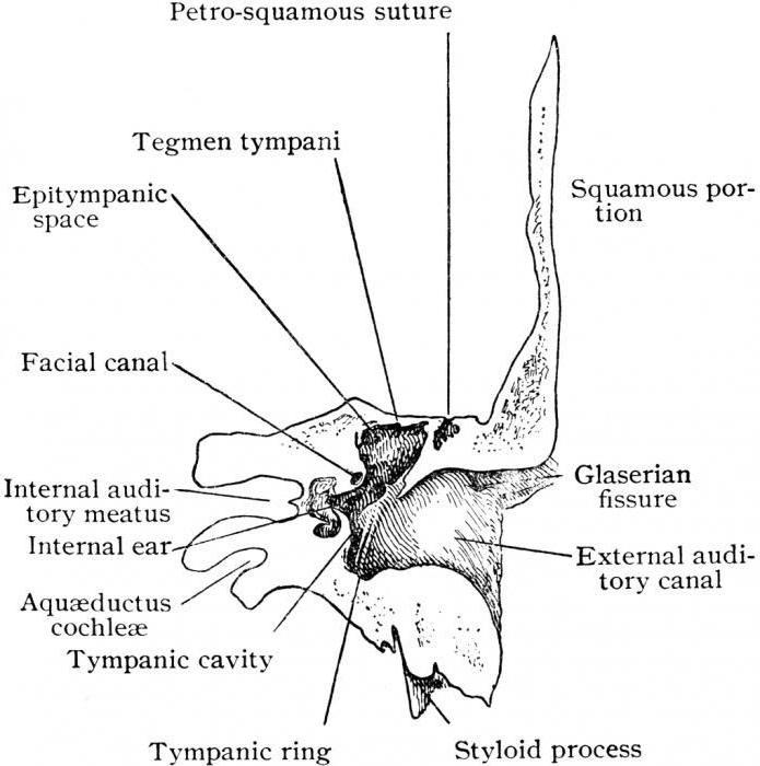
The tympanic part (pars tympanica) is fused with the mastoid process (processus mastoideus) and the scaly part (pars squamosa), is a thin plate that bounds the external auditory opening (porus acusticus externus) and the external auditory canal (meatus acusticus externus) in front, behind and below. .
The stony part (pars petrosa) has the shape of a three-sided pyramid, the apex of which faces anteriorly and medially, and the base, which passes into the mastoid process (processus mastoideus), faces posteriorly and laterally.
There are three surfaces: anterior, posterior and lower, as well as three edges: anterior, posterior and upper.
The anterior surface (facies anterior) is part of the bottom of the middle cranial fossa; posterior (facies posterior) faces backward and medially, forms part of the anterior wall of the posterior cranial fossa; the lower one (facies inferior) faces down and is visible only on the outer surface of the base of the skull.
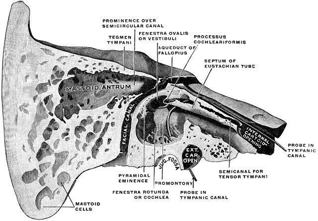
The external relief of the pyramid is due to its structure as a container for the middle and inner ear, as well as for the passage of blood vessels and nerves.
A thin pointed styloid process (processus styloideus) extends from the lower surface of the pyramid, serving as a site of muscle attachment. The relief of the outer surface of the pyramid is the site of muscle attachment; downward it extends into the mastoid process, to which the sternocleidomastoid muscle is attached.
On the mastoid process (on its anterior smooth surface) of the temporal bone, the Shipo triangle is distinguished, which is the site of operational access to the cells of the mastoid process. On an x-ray of the temporal bones, the so-called sinodural angle (Citelli's angle) is distinguished. Inside, the mastoid process contains cells (cellulae mastoideae), which are air cavities communicating with the tympanic cavity (middle ear) through the mastoid cave (antrum mastoideum).
Temporal bone connected to the occipital, parietal and sphenoid bones. Participates in the formation of the jugular foramen.
Temporal bone canals
- sleepy channel, canalis caroticus, in which the internal carotid artery lies. It begins on the lower surface of the pyramid, with the external carotid foramen (foramen caroticum externum), directed vertically upward, bending at a right angle, directed forward and medially. The canal opens into the cranial cavity through the internal carotid foramen (foramen caroticum internum).
- drum string channel, canaliculus chordae tympani, starts from the canal facial nerve, slightly above the stylomastoid foramen (foramen stylomastoideum), goes forward and opens into tympanic cavity. A branch of the facial nerve, the chorda tympani, passes through this canaliculus, which then exits the tympanic cavity through the petrotympanic fissure (fissura petrotympanica).
- facial canal, canalis facialis, in which the facial nerve passes, it begins at the bottom of the internal auditory canal, then runs horizontally from back to front. Having reached the level of the cleft of the canal of the greater petrosal nerve, the canal goes back and laterally, at a right angle, forming a bend, or elbow, of the facial canal. Next, the channel is directed back, following horizontally along the axis of the pyramid. Then it turns vertically downwards, bending around the tympanic cavity, and on the lower surface of the pyramid it ends with a stylomastoid opening.
- myotubal canal, canalis musculotubaris, has a common wall with the carotid canal. It begins in the angle formed by the anterior edge of the pyramid and the squama of the temporal bone, running posteriorly and laterally, parallel to the anterior edge of the pyramid. The muscular-tubal canal is divided into two half-canals by a longitudinal horizontal septum. The upper hemicanal is occupied by the tensor tympani muscle, and the lower is the bony part of the auditory tube. Both canals open into the tympanic cavity on its anterior wall.
- mastoid tubule, canaliculus mastoideus, originates at the bottom of the jugular fossa and ends in the tympanomastoid fissure. A branch of the vagus nerve passes through this canaliculus.
- tympanic canaliculus, canaliculus tympanicus, arises in the stony fossa (fossula petrosa) with an opening through which a branch of the glossopharyngeal nerve, the tympanic nerve, enters. After passing through the tympanic cavity, this nerve, called the lesser petrosal nerve, exits through the cleft of the same name on the anterior surface of the pyramid.
- carotid tympanic tubules, canaliculi caroticotympanici, pass in the wall of the internal carotid artery canal near its external opening and open into the tympanic cavity. They serve for the passage of the vessels and nerves of the same name.
- vestibule water supply, aqueductus vestibuli, a canal in the pyramid of the temporal bone that connects the vestibule of the bony labyrinth (the expanded part of the bony labyrinth between the cochlea of the inner ear and the bony semicircular canals) with the cranial cavity (posterior cranial fossa). It opens with a gap on the posterior surface of the pyramid of the temporal bone, behind the opening of the internal auditory canal. The canal contains the vein of the aqueduct of the vestibule and the ductus endolymphaticus, which ends in the blind sac (saccus endolymphaticus), on the posterior surface of the pyramid of the temporal bone, between the opening of the internal auditory canal and the sigmoid sinus.
- snail plumbing, aqueductus cochleae, about 10 mm long, connects the vestibule of the inner ear and the posterior surface of the pyramid of the temporal bone, opening at its lower edge, below the opening of the internal auditory canal. Its internal opening is located at the beginning of the staircase of the drum of the bony cochlea. The vein of the cochlear canaliculus passes through the canal.
The temporal bone, the anatomy of which will be discussed further, is a pair. It contains organs of balance and hearing. The temporal bone of the skull takes part in the formation of its base and lateral wall of the vault. Articulating with the lower jaw, it provides support for the masticatory apparatus. Next, let's take a closer look at what the temporal bone is.
Anatomy
There is an auditory opening on the outer surface of the element. There are three parts around it: scaly (above), petrous (or pyramid of the temporal bone) - behind and inward, tympanic - below and in front. The rocky area, in turn, has 3 surfaces and the same number of edges. The left and right temporal bones are the same. The segments contain channels and cavities.
Scaly part
It is presented in the form of a plate. The outer surface of this part is slightly rough and has a slightly convex shape. In the posterior section, the groove of the temporal (middle) artery runs in a vertical direction. An arcuate line runs along the posterior lower section. From the scaly part, the zygomatic process extends slightly anteriorly and from above in a horizontal direction. It seems to be a continuation of the ridge located on the outer surface along the lower edge. Its beginning is presented in the form broad root. Then the process narrows. It has an outer and inner surface and 2 edges. One, the top one, is longer, and the second, the bottom one, is correspondingly shorter. The front end of the element is jagged. The processes of the temporal bone in this area are connected using a suture. As a result, the zygomatic arch is formed. The mandibular fossa is located on the lower surface of the root. It has a transverse oval shape. The anterior part of the fossa - half up to the stony-squamous fissure - is articular surface temporomandibular joint. In front, the fossa is limited by a tubercle. The outer plane of the scaly part takes part in the formation of the temporal fossa. This is where the muscle bundles originate. On the inner surface there are finger-like impressions and an arterial groove. The latter contains the meningeal (middle) artery. 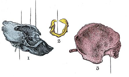
The edges of the scaly part
There are two of them: parietal and sphenoid. The latter - jagged and wide - articulates with the squamosal edge in the large wing of the sphenoid bone. As a result, a seam is formed. The upper posterior parietal edge is longer than the previous one, pointed and articulates with the squamosal in the parietal bone.
Rocky part
The structure of the temporal bone in this area is quite complex. The petrous part includes the anteromedial and posterolateral sections. The latter is the mastoid process of the temporal bone. It is located posterior to the auditory (external) opening. It distinguishes between internal and external surfaces. The outer one is rough and has a convex shape. Muscles are attached to it. Inferiorly the process becomes a protrusion. It has a cone shape and can be felt quite well through skin. WITH inside there is a deep notch. Parallel to it and slightly posteriorly there is a groove of the occipital artery. The posterior border of the process is the occipital jagged edge. Connecting, the edges in this area form a seam. In the middle of its length or at the occipital end there is a mastoid foramen. In some cases there may be several. The emissary mastoid veins lie here. From above, the process is limited by the parietal edge. At the border with the area of the scaly part of the same name, it forms a notch. The angle from the parietal bone enters it and forms a suture. 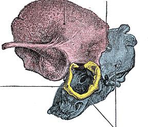
Rocky surfaces
There are three of them. The anterior surface is wide and smooth. It faces the cranial cavity, is directed obliquely forward and from top to bottom, and passes into the medullary plane of the scaly part. Almost in the center on the front surface there is an arched elevation. It is formed by the semicircular anterior canal of the labyrinth, which lies below. Between the gap and the elevation there is a roof of the drum part. The posterior surface of the stony part, like the anterior one, turns into the cranial cavity. However, it is directed posteriorly and upward. The posterior surface is continued by the mastoid process. Almost in the middle there is an auditory (internal) opening leading to the corresponding passage. The lower surface is uneven and rough. It forms part of the lower plane of the base of the skull. There is an oval or round jugular fossa. At its bottom there is a small groove leading to the opening of the mastoid canaliculus. The posterior edge of the fossa is limited by the notch. It is divided into two parts by a small process. 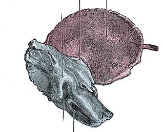
Edges of rocky area
There is a groove in the upper edge of the pyramid. It represents the imprint of the venous sinus lying here and the fixation of the tentorium of the cerebellum. The posterior edge of the rocky area separates the posterior and inferior surfaces. The petrosal sinus groove runs along the brain surface along it. Almost in the middle of the posterior edge, near the jugular notch, there is a funnel-shaped triangular depression. The anterior edge is shorter than the posterior and upper edge. It is separated from the scaly part by a gap. At the anterior edge there is an opening of the muscular-tubal canal leading to the tympanic cavity.
Channels of the rocky part
There are several of them. The carotid canal originates in the middle sections on the lower surface in the stony part with an external opening. At first it is directed upward. Further, bending, the canal follows medially and anteriorly, opening at the apex of the pyramid with an opening. The carotid tympanic tubules are small branches. They lead into the tympanic cavity. The facial canal begins at the bottom of the internal auditory canal. It runs horizontally and almost at a right angle relative to the axis of the stony section. Next, the channel is directed to the front surface. In this place, turning at an angle of 90 degrees, it forms a knee. Next, the canal passes to the posterior part of the medial wall in the tympanic cavity. Then, heading posteriorly, it passes along the axis in the rocky part until it reaches an eminence. From this place it goes down vertically, opening with a stylomastoid foramen. 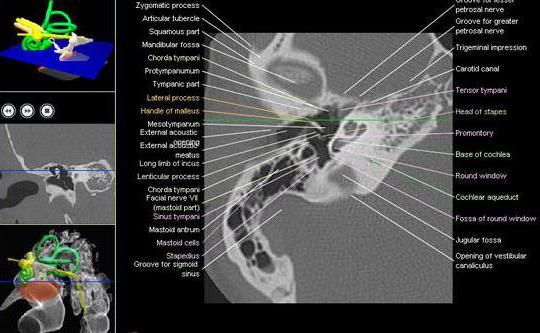
Drum string channel
It begins a few millimeters higher than the stylomastoid foramen. The channel goes up and forward, entering the tympanic cavity and opening on its back wall. The chorda tympani, a branch of the intermediate nerve, passes through the canaliculus. It exits the cavity through the petrotympanic fissure.
Musculo-tubal canal
It is a continuation of the anterior superior region of the tympanic cavity. Its external opening is located near the notch between the scaly and petrous parts of the bone. The canal runs laterally and somewhat posteriorly from the horizontal section of the carotid tract, almost along the longitudinal axis of the petrous section. There is a partition inside it. It is located horizontally. By means of this partition the canal is divided into two parts. The upper one is the semi-canal of the muscle that strains the eardrum. The large inferior section belongs to the auditory tube.
Tympanic canaliculus
It starts from the lower surface in the pyramidal part, in the depths of the rocky pit. Further it is directed towards lower cavity, perforating which, it passes along the medial wall, reaching the groove of the promontory. Then he goes to the top plane. There it opens into a cleft in the petrosal nerve canal. 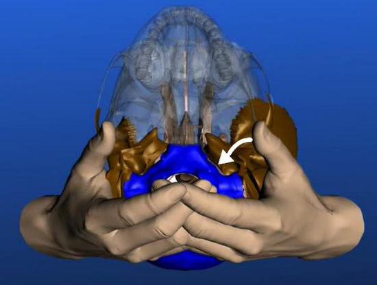
Drum part
This is the smallest section that includes the temporal bone of the skull. It is presented in the form of a slightly curved ring-shaped plate. The tympanic part forms part of the posterior, inferior and anterior walls of the auditory canal (external meatus). Here you can see the border fissure, which, together with the stony fissure, delimits this area from the mandibular fossa. The outer edge is closed at the top by bone scales. It delimits the auditory (external) opening. There is a spine at its posterior upper outer edge. Below it is the supraductal fossa.
Damage
One of the most serious injuries is a fracture of the temporal bone. It can be either longitudinal or transverse. Both types of injury, unlike injuries to other bones, are characterized by the absence of movement of fragments. Due to this, the width of the gap is usually small. An exception is impression damage to the scales. In such cases, quite significant displacement of the fragments may be observed.
CT scan of the temporal bones
The study is used when there are suspected violations in the structure of an element. Computer diagnostics is a special method. With its help, the temporal bone is scanned layer by layer. This creates a series of images. The temporal bone is examined in cases of the presence of:
- Injuries on one or both sides.
- Otitis, especially of unknown nature.
- Balance and hearing disorders, signs of dysfunction of the formations next to which the temporal bone is located.
- Otosclerosis.
- Suspected tumor in structures located near or inside the temporal bone.
- Mastoiditis.
- Brain abscess in close proximity to the bone.
- Discharge from the ear.
Tomography of the temporal bones is also indicated in preparation for electrode implantation. 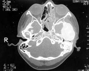
Contraindications for the study
Computed tomography allows specialists to obtain accurate information about the condition of the temporal bones and is considered one of the best diagnostic methods for various disorders. However, in some cases it is necessary to refuse this procedure. This is due to the presence of contraindications in patients. Among them it should be noted:
- All stages of pregnancy. Impact ionizing radiation, generated by the tubes of the device, can provoke the development of fetal pathology.
- Overweight. Structurally, the tomograph is not intended for examining obese patients.
- Increased sensitivity to contrast agents. When a compound is introduced into the body, severe allergic reaction, up to anaphylactic shock.
- Kidney failure. In this case, patients contrast agent is not excreted from the body, which can cause harm to health.
There are other limitations to diagnostics. They are quite rare.
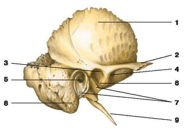
The temporal bone (os temporale) is paired; it contains the organs of hearing and balance. Nerves and blood vessels pass through its channels. The bone consists of three parts (Fig. 51).
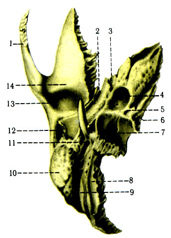
The scales (squama) have the shape of an oval thin plate located vertically, almost in the sagittal plane. The zygomatic process (processus zygomaticus) begins from the temporal surface of the scales. At the beginning of this process, on the lower surface of the scales, there is a mandibular fossa (fossa mandibularis), in front of which there is an articular tubercle (tuberculum articulare). On the cerebral surface of the scales there are imprints from the middle meningeal artery (a. meningea media) and the convolutions of the temporal lobe of the brain.
The tympanic part (pars tympanica) has the shape of a semiring and takes part in the construction of the anterior, lower and posterior walls of the external auditory canal (meatus acusticus externus), the upper wall of which is limited by scales.
The stony part (pyramid) (pars petrosa) is triangular in shape, facing medially and anteriorly, has anterior, posterior and inferior surfaces, anterior, superior and posterior edges.
On the front surface of the stony part, when connecting it to the scales, there is a platform - the roof of the tympanic cavity (tegmen tympani). In front, this area is limited by a fissure (fissura petrosquamosa), and laterally by an arcuate elevation (eminentia arcuata). Below it are the anterior and posterior semicircular canals of the inner ear. From the eminentia arcuata, closer to the apex of the pyramid, there are two openings representing the exit points of the greater and lesser petrosal nerves (hiatus canalis n. petrosi majoris et minoris), opening into the grooves of the same name, which are oriented towards the apex of the pyramid.
On the posterior surface of the petrous part there is an internal auditory opening (porus acusticus internus), where the facial and vestibulocochlear nerves pass. At the base of the stony part there is a deep sigmoid groove (sulcus sigmoideus), into which the opening of the mastoid venous outlet opens. Lateral to the internal auditory canal there is a slit-like opening of the aqueduct of the vestibule of the inner ear (apertura externa aqueductus vestibuli). On the upper edge, between the anterior and posterior surfaces of the stony part, there is a groove (sulcus sinus petrosi superioris), which reaches the sigmoid groove behind and the apex of the pyramid in front.
On the lower surface of the base of the stony part there is a styloid process (processus styloideus); behind it, the stylomastoid foramen (for. stylomastoideum) opens, representing the opening of the facial nerve canal. More medially styloid process the jugular fossa (fossa jugularis) is visible, the posterior edge of which has a notch of the same name. The anterior edge of the jugular fossa borders the external opening of the carotid canal (for. caroticum externum). In the anterior edge there is a small stony fossa (fossula petrosa), at the bottom of which the tympanic canal (canaliculus tympanicus) begins. In adults, behind the stylomastoid foramen and the external auditory canal is the mastoid process (processus mastoideus). In its thickness there are cells lined with mucous membrane and communicating with the tympanic cavity. Medial to the mastoid process are the mastoid fissure and the occipital groove. The latter contains the occipital artery. In the middle of the posterior edge of the pyramid there is an external opening of the cochlea's aqueduct (apertura externa canaliculi cochleae).
Temporal bone canals. The carotid canal (canalis caroticus) begins on the lower surface of the pyramid with the external opening of the same name. The channel in the thickness of the pyramid turns at an angle of 90° and goes to the top of the pyramid, where it ends with an internal opening (for. caroticum internum).
The facial canal (canalis facialis) begins in the internal auditory canal, then crosses the pyramid transversely and at the cleft of the greater petrosal nerve (hiatus canalis n. petrosi majoris) turns at a right angle to side- the knee of the facial canal (geniculum canalis facialis), then goes laterally, located at the junction of the roof of the tympanic cavity with the labyrinthine wall of the inner ear. At the posterior wall of the tympanic cavity, the canal turns and goes down, ending on the lower surface of the pyramid of the temporal bone with a stylomastoid foramen.
The muscular-tubal canal (canalis musculotubarius) is limited by the anterior edge of the apex of the pyramid and the scales. It consists of two sections: half-channel auditory tube(semicanalis tubae auditivae) and the hemicanal of the muscle that stretches the tympanic membrane (semicanalis m. tensoris tympani).
The tympanic canaliculus (canaliculus tympanicus) is very narrow; begins in the fossula petrosa and opens on the anterior surface of the petrous part of the pyramid with the cleft of the canal of the lesser petrosal nerve (hiatus canalis n. petrosi minoris).
The canaliculus chordae tympani extends from the facial canal before it exits the petrous part. It opens into the petrotympanic fissure of the mandibular fossa.
Ossification. The temporal bone of a newborn consists of three independent parts, which are described above. The external auditory canal is relatively short and wide. The tympanic cavity is filled with loose connective tissue, which resolves within the first 3 months. after birth.
The tympanic part is presented in the form of an incomplete ring located under the scales lateral to the pyramid. In the lumen of the ring is stretched eardrum. The process of ossification occurs in connective tissue (primary bone), bypassing the cartilaginous stage. By the age of 6 years, the external auditory canal develops from the semiring, scales and mastoid process. At the 8th week of intrauterine development, three ossification points appear in the fibrous connective tissue of the scales. From the back of the scales and the lateral part of the pyramid, under the action of the sternocleidomastoid muscle, the mastoid process is formed, which is pneumatized in three stages: up to 1 year, a tympanic invagination is formed, up to 3 years, cells are formed, and up to 6 years, pneumatization of the process is completely completed. In the cartilaginous base of the pyramid for V months. During intrauterine development, 5 bone nuclei appear, which merge at the time of birth.
Full text search:
Home > Abstract >Medicine, health
Ministry of the Republic of Kazakhstan
Karaganda State Medical University
SRSP
The structure of the temporal and sphenoid bones. Canals of the temporal bone.
Completed by: student Gabdolla I.K.
Teacher: Harutyunyan M.G.
Karaganda - 2011
Temporal bone (os temporale) steam room, part of the base and side wall of the skull between the sphenoid bone in front and the occipital bone in the back. It houses the organs of hearing and balance. The temporal bone is divided into pyramid, tympanic and squamosal parts.
Pyramid, or rocky part(pars petrosa), has a triangular shape, located obliquely in the horizontal plane. The apex of the pyramid is directed forward and medially, and the base is directed backward and laterally. At the top of the pyramid is the internal opening of the carotid canal (canalis caroticus). Nearby and to the lateral side is the muscular-tubal canal (canalis musculotubarius), which is divided by a septum into two semi-canals: the semi-canal of the auditory tube (semicanalis tubae auditivae) and the semi-canal of the tensor tympani muscle (semicanalis musculi tensoris tympani).
The pyramid has three surfaces: front, back and bottom. Front surface The pyramid faces up and forward. Near the apex on this surface there is a small trigeminal impression (impressio trigemini). Two openings are visible lateral to this depression. The larger of them is called the cleft (hole) of the canal of the greater petrosal nerve (hiatus canalis nervi petrosi majoris), from which a narrow groove of the same name runs forward and medially. Anterior and lateral is the cleft of the lesser petrosal nerve (hiatus canalis nervi petrosi minoris), which passes into the groove of this nerve. On the front surface of the pyramid there is a flattened section - the roof of the tympanic cavity (tegmen thympani), which is its upper wall. Along the upper edge of the pyramid there is a groove of the superior petrosal sinus (sulcus sinus petrosi superioris).
Back surface of the pyramid facing posteriorly and medially. In the middle of this surface is the internal auditory opening (porus acusticus internus). It leads into the internal auditory canal (medtus acusticus internus). Lateral and slightly above this opening is the subarc fossa (fossa subarcuata), below and lateral to which there is a little noticeable external aperture (hole) of the vestibular aqueduct (apertura externa aqueductus vestibuli). The groove of the inferior petrosal sinus (sulcus sinus petrosi inferioris) runs along the posterior edge of the pyramid. At the lateral end of this groove, adjacent to the jugular fossa, there is a depression, at the bottom of which the external aperture of the cochlear canaliculus (apertura externa canaliculi cochleae) opens.
Bottom surface of the pyramid has a complex terrain. Near the base of the pyramid there is a deep jugular fossa (fossa jugularis). Anterior to it there is a rounded external opening of the carotid canal, inside of which, in its wall, there are 2-3 openings of the carotid-tympanic tubules connecting the carotid canal with the tympanic cavity. On the ridge between the jugular fossa and the external opening of the carotid canal there is a small lobe (fossula petrosa). Lateral to the jugular fossa, a thin and long styloid process (processus styloideus) is directed downwards. Behind the process there is a stylomastoid foramen (foramen stylomastoideum), and behind this opening a wide, easily palpable mastoid process (processus mastoideus) is directed downwards.
In the thickness of the mastoid process there are air-filled cells. The largest cell, the mastoid cave (Antrum mastoideum), communicates with the tympanic cavity. Medially, the mastoid process is limited by the deep mastoid notch (incisure mastoidea). Medial to this notch is the groove of the occipital artery (sulcus arteriae occipitalis). At the base of the mastoid process there is sometimes a mastoid foramen (foramen mastoideum).
Drum part(pars tympanica) is formed by a curved narrow bone plate, which in front, below and behind limits the external auditory opening (porus acusticus externus), leading into the external auditory canal (meatus acusticus externus). Between drum part and the mastoid process there is a narrow tympanomastoid fissure (fissure tympanomastoidea). In front of the external auditory opening is the tympanic-squamous fissure (fissure tympanosquamosa). A narrow bone plate protrudes into this gap from the inside - the edge of the roof of the tympanic cavity. As a result, the tympanic-squamous fissure is divided into the anterior stony-squamous fissure (fissura petrosquamosa) and the petrotympanic fissure (fissura petrotympanica, Glaser's fissure), through which a branch of the facial nerve, the chorda tympani, emerges from the tympanic cavity.
Scaly part(pars squamosa) is a convex plate outward, having a beveled free upper edge for connection with the parietal bone and the greater wing of the sphenoid bone. The outer temporal surface of the scales is smooth. On the inner cerebral surface of the scales there are cerebral elevations, finger-like impressions and arterial grooves. From the scales, above and anterior to the external auditory canal, the zygomatic process (processus zygomaticus) begins. Connecting with temporal process zygomatic bone, it forms the zygomatic arch. Behind the zygomatic process, at its base, is the mandibular fossa (fossa mandibularis) for articulation with the condylar process of the mandible to form the temporomandibular joint.
Canals of the temporal bone.
Sleepy channel canalis cardticus) begins on the lower surface of the pyramid with the external carotid foramen, goes upward, bends almost at a right angle, then goes medially and forward. The canal ends with the internal carotid foramen at the top of the pyramid of the temporal bone. The internal carotid artery and the nerves of the carotid plexus pass through this canal into the cranial cavity.
Carotid tympanic tubules(canaliculi caroticotympanic!), numbering 2-3, depart from the carotid canal and go to the tympanic cavity. These tubules contain arteries and nerves of the same name.
Musculo-tubal canal(canalis musculotubarius) begins at the top of the pyramid of the temporal bone, goes back and laterally and opens into the tympanic cavity. A horizontal partition divides it into two parts. Above is the hemicanal of the tensor tympani muscle (semicanalis musculi tensoris tympani), containing the muscle of the same name. Below is the semicanal of the auditory tube (semicanalis tubae auditivae).
Facial canal(canalis facialis) begins in the internal auditory canal. It first runs transverse to the long axis of the pyramid to the level of the cleft of the greater petrosal nerve canal. Having reached the cleft, the canal forms a knee, then is directed at a right angle back and laterally. Having passed along the medial wall of the tympanic cavity, the canal turns vertically downwards and ends with the stylomastoid foramen. The facial nerve passes through this canal.
Drum string channel(canaliculus chordae tympani) comes from the wall of the facial canal in its final section and opens into the tympanic cavity. The nerve, the chorda tympani, passes through this canal.
Drum tubule(canaliculus tympanicus) begins at the bottom of the stony dimple, goes up, pierces the wall of the tympanic cavity. The canaliculus then passes along its medial wall and ends in the area of the cleft of the canal of the lesser petrosal nerve. The tympanic nerve passes through this canaliculus.
Mastoid tubule(canaliculus mastoideus) begins in the jugular fossa and ends in the tympanomastoid fissure. The auricular branch of the vagus nerve passes through this canaliculus.
Several channels of the temporal bone pass through the pyramid for cranial nerves and blood vessels.
Sleepy channel(canalis caroticus) begins on the lower surface of the pyramid with the external carotid foramen, goes upward, bends almost at a right angle, then goes medially and forward. The canal ends with the internal carotid foramen at the top of the pyramid of the temporal bone. The internal carotid artery and the nerves of the carotid plexus pass through this canal into the cranial cavity.
Carotid tympanic tubules(canaliculi caroticotympanici), numbering 2-3, depart from the carotid canal and go to the tympanic cavity. These tubules contain arteries and nerves of the same name.
Musculo-tubal canal(canalis musculotubularis) begins at the top of the pyramid of the temporal bone, goes back and laterally and opens into the tympanic cavity. A horizontal partition divides it into two parts. Above is the hemicanal of the tensor tympani muscle (semicanalis musculi tensoris tympani), containing the muscle of the same name. Below is the semicanal of the auditory tube (semicanalis tubae auditivae).
Facial canal(canalis facialis) begins in the internal auditory canal. It first runs transverse to the long axis of the pyramid to the level of the cleft of the canal of the greater petrosal nerve. Having reached the cleft, the canal forms a knee, then is directed at a right angle back and laterally. Having passed along the medial wall of the tympanic cavity, the canal turns vertically downwards and ends with the stylomastoid foramen. The facial nerve passes through this canal.
Drum string channel(canaliculus chordae tympani) comes from the wall of the facial canal in its final section and opens into the tympanic cavity. The nerve, the chorda tympani, passes through this canal.
Tympanic canaliculus(canaliculus tympanicus) begins at the bottom of the stony dimple, goes up, pierces the wall of the tympanic cavity. The canaliculus then passes along its medial wall and ends in the area of the cleft of the canal of the lesser petrosal nerve. The tympanic nerve passes through this canaliculus.
Mastoid tubule(canaliculus mastoideus) begins in the jugular fossa and ends in the tympanomastoid fissure. The auricular branch of the vagus nerve passes through this canaliculus.
Sphenoid bone (os sphenoidale) occupies a central position at the base of the skull. It participates in the formation of the base of the skull, its lateral sections and a number of cavities and pits. The sphenoid bone consists of a body, pterygoid processes, greater and lesser wings.
Body of the sphenoid bone(corpus sphenoidale) has an irregular shape and six surfaces: upper, lower, posterior, fused (in an adult) with the basilar part of the occipital bone, anterior and two lateral surfaces. On the upper surface of the body there is a depression - the sella turcica (sella turcica) with a deep pituitary fossa (fossa hypophysialis). At the back of the sella turcica there is a dorsum sellae (dorsum sellae), and at the front there is a tuberculum sellae (tuberculum sellae). On each side of the body of the bone, a carotid groove (sulcus caroticus) is visible - a trace of the attachment of the internal carotid artery. On the anterior surface of the body of the sphenoid bone there is a wedge-shaped crest (crista sphenoidalis). On the sides of the crest there are irregularly shaped wedge-shaped shells (conchae sphenoidales), limiting the apertures of the sphenoid sinus. The sphenoid sinus (sinus sphenoidalis) is an air-filled cavity communicating with the nasal cavity.
The lateral surfaces of the body of the sphenoid bone directly pass into paired small and large wings.
Small wing(ala minor) is a laterally directed flattened bone plate, at the base of which is the optic canal (canalis opticus), leading to the orbit. The posterior free edge serves as the boundary between the anterior and posterior cranial fossae. The anterior margin connects with the orbital part of the frontal bone and the cribriform plate of the ethmoid bone. Between the small wing at the top and the upper edge big wing there is an elongated opening - the superior orbital fissure (fissura orbitalis superior), connecting the cranial cavity with the orbit.
Big wing(ala major) starts from the lateral surface of the body of the sphenoid bone with a wide base and, like the lesser wing, is directed to the lateral side. It has four surfaces: medullary, orbital, temporal and maxillary. The concave surface of the brain faces the cranial cavity. It has three holes through which blood vessels and nerves pass. A round opening (foramen rotundum), located closer to the base of the greater wing, leads into the pterygopalatine fossa. At the level of the middle of the wing there is an oval foramen (foramen ovale), which opens at the base of the skull, and behind it there is a small spinous foramen (foramen spinosum). The orbital surface (facies orbitalis) is smooth and participates in the formation of the lateral wall of the orbit. On the temporal surface (facies temporalis) there is an infratemporal crest (crista infratemporalis), oriented in the anteroposterior direction and delimiting the temporal fossa from the infratemporal fossa on the lateral surface of the skull.
The maxillary surface (facies maxillaris) faces forward - into the pterygopalatine fossa.
Pterygoid process(processus pterygoideus) paired, extends downwards from the body of the sphenoid bone. The process consists of medial and lateral plates (lamina medialis et lamina lateralis). At the back between the plates there is a pterygoid fossa (fossa pterygoidea). At the base of the pterygoid process, a narrow pterygoid canal (canalis pterygoideus) runs from back to front, connecting the pterygopalatine fossa with the area of the foramen lacerum on the whole skull.
Occipital bone (os occipitale) is located in the posterior lower part of the cerebral part of the skull. This bone contains a basilar part, two lateral parts and an occipital scale, which surround the large (occipital) foramen (foramen magnum).
Basilar part(pars basilaris) is located in front of the large (occipital) foramen. In front, it connects with the body of the sphenoid bone, together with which it forms a platform - the slope (clivus). On the lower surface of the basilar part there is an elevation - the pharyngeal tubercle (tuberculum pharyngeum), and along the lateral edge there is groove of the inferior petrosal sinus(sulcus sinus petrosi inferioris).
Lateral part(pars lateralis) steam room, from behind it passes into the scales of the occipital bone. Below on each lateral part there is an ellipsoidal elevation - the occipital condyle (condylus occipitalis), at the base of which is the canal of the hypoglossal nerve (canalis nervi hypoglossi). Behind the condyle there is a condylar fossa (fossa condylaris), and at its bottom there is an opening of the condylar canal (canalis condylaris). On the side of the occipital condyle is the jugular notch (incisura jugularis), which, together with the jugular notch of the pyramid of the temporal bone, forms the jugular foramen. Next to the jugular notch on the brain surface there is a groove for the sigmoid sinus (sulcus sinus sigmoidei).
Occipital scales(squama occipitalis) - a wide, convex plate outward, the edges of which are highly jagged. On the whole skull they connect to the parietal and temporal bones. In the center of the outer surface of the scales, the external occipital protuberance (protuberantia occipitalis externa) is visible, from which a weakly defined upper line (linea nuchae superior) extends in both directions. The external occipital crest (crista occipitalis externa) runs down from the protrusion to the foramen magnum (occipital foramen). From its middle, a lower line (hinea nuchae inferior) goes to the right and left. The highest line (linea nuchae suprema) is sometimes visible above the external occipital protrusion.
On the inner side of the occipital scales there is a cruciform eminence (eminentia cruciformis), dividing the brain surface of the scales into 4 pits. The center of the cruciform eminence forms the internal occipital protuberance (protuberantia occipitalis interna). To the right and left of this protrusion there is a groove of the transverse sinus (sulcus sinus transversus). Up from the protrusion there is a groove of the superior sagittal sinus (sulcus sinus sagittalis superioris), and down, to the large (occipital) foramen, there is the internal occipital crest (crista occipitalis interna).
Bones (Fig. 4, 2) lies a small main wedge-shaped bone(basisphenoideum). It is completely covered by a wide main temporal bone... shock-absorbing device - forks), structure wing skeleton (including...
Every bone human body is the most important “cog” in a huge mechanism. The head bone elements perform protective function. These elements include the temporal bone.
Temporal bone: description
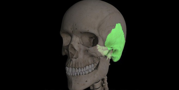
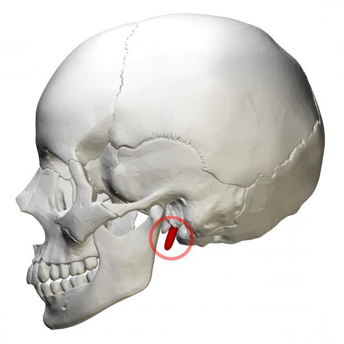
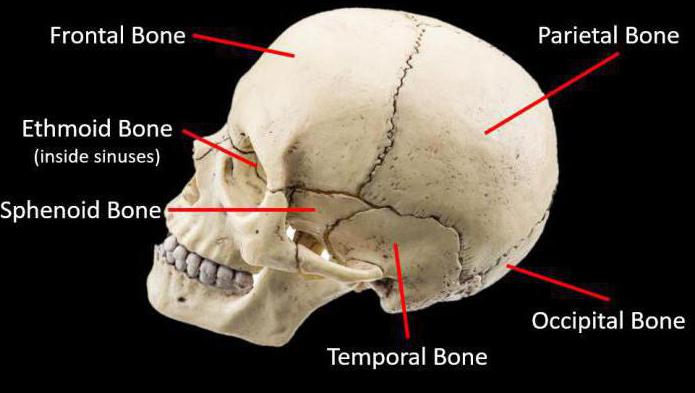
An important part of the skull is the temporal bone, which is located on both sides of the skull, and therefore is a pair. More precisely, it belongs to one of the components of the skull that covers the brain. It is surrounded by the sphenoid, parietal and occipital bones.
This bone element in combination with lower jaw forms a movable joint. And in tandem they form the zygomatic arch.
The temporal element itself is not a single bone: it is represented by a number of parts that form it.
The temporal bone develops by ossification from six points. At the end of the 8th week of embryonic development, the scaly parts are the first to ossify. In the 3rd month, hardening occurs in the tympanic part. With the arrival of the 5th month of fetal development, several areas of ossification appear in the cartilaginous portion of the pyramid.
By the period preceding birth, the temporal bone already consists of a scaly part, a tympanic and a petrous part, and in between these parts there are clefts with connective tissue.
Bone structure
The anatomy of the temporal bone is as follows. It consists of a pyramid, a drum part and scales.
The pyramid is also called the rocky part. And for good reason, because this element consists of a very hard bone element. According to its form rocky part very similar to a three-sided pyramid (hence the name). The base of the pyramid extends into the mastoid process.
The pyramid consists of the following parts: the top; front, back and bottom surfaces; apical, posterior and inferior margins.
The frontal surface of the pyramid is directed forward and upward. On the lateral side, the pyramid passes into the scales of the temporal bone. Between these two elements of the temporal bone is the petrosquamosal foramen. In its central part, the front surface of the pyramid has a small arched elevation. At a distance through these elevations, in the form of a scaly hole, there is a flat section that serves as a roof.
The back surface of the pyramid is adjacent to the center. Almost in the central part of this surface of the pyramid there is a small auditory opening, which flows into the internal auditory canal. On the lateral side of the auditory opening is the subarcular fossa. And according to bottom side there is an opening for the vestibule water supply.
The lower surface of the pyramid is equipped with a complex surface relief. The lower surface flows into the mastoid process.
The upper edge of the pyramid is the boundary line connecting the front and rear surfaces. At its base there is a groove for the petrosal sinus.
The posterior edge of the pyramid separates the posterior and inferior surfaces. Along its surface lies the groove of the inferior petrosal sinus. Near the side of the groove there is a dimple with the external opening of the cochlear canaliculus.
On the inside, the pyramid houses the organs of hearing and balancing.
The diagram shows:
Functions
The temporal bone has three functions:
- Protective. The temporal bone, together with the rest of the bones of the skull, protects the brain from various types of damage.
- Support. The cranial bone supports the brain, being its support.
- The temporal bone is the attachment point for the head muscles.
In addition, this bone contains organs and canals hearing aid, equilibrium, and also contains various tubules and vessels.
The functions performed depend entirely on the anatomy of the temporal bone. Additionally, the location of nearby bones also affects functionality.
Temporal bone canals
The temporal bone is completely striated with various notches, depressions and canaliculi. The canals and cavities of the temporal bone serve to conduct blood vessels, nerve branches, and arteries. The canals are hollow tubular cords that intertwine parts of the temporal bone.
Below is a table of the temporal bone canals.
| Bone canals | What cavities connect | What crosses the channels |
| Facial canal | Dorsal wall of the pyramid and stylomastoid foramen | 7th petrosal artery and stylomastoid vessels |
| Sleepy channel | Apex of the pyramid and outer base of the skull | Carotid artery and carotid plexus |
| Musculo-tubal canal | Tympanum and superior wall of the pyramid | Superior tympanic artery, auditory tube |
| Drum string channel | Facial canal, tympanic cavity and tympanic fissure | 7th facial nerve and posterior tympanic artery |
| Mastoid tubule | Jugular recess and mastoid fissure | auricular process of the 10th pulmonary gastric nerve |
| Tympanic canaliculus | Petrosal fossa, inferior wall of the pyramid and tympanic cavity | Small stony nerve vessel, tympanic artery lying inferiorly |
| Carotid tympanic tubules | The edge of the carotid cord and the tympanic cavity | Carotid-tympanic nerve fibers and arteries |
| Snail tubule | The beginning of the internal auditory organ and the lower base of the pyramid | Cochlear canaliculus vein |
| Internal auditory canal | Inner ear and posterior cranial fossa | 7th facial nerve, 8th cochlear nerve and inner ear artery |
| Plumbing vestibule | The beginning of the inner ear and the cranial fossa located on the back side | Venous vessel of the aqueduct |
Facial nerve canal
Let's look at the facial canal of the temporal bone. It originates on the underside of the hearing aid, located inside the ear. Its direction is expressed laterally - forward to the cleft of the petrosal nerve fiber canal. In this area it forms a turn, which is called the elbow of the facial canal. The facial canal of the temporal bone continues its path from the knee in the direction of the side and back, along the trajectory of a right angle parallel to the axis of the pyramid. Then the direction becomes vertical and ends with the mastoid opening at the rear wall of the tympanic cavity.
Sleepy channel
The carotid canal of the temporal bone begins its journey on the underside of the pyramid in the form of a hole (aperture). Its direction is straight and upward, but closer to the surface of the pyramid. The canal bends at an angle of 90 and exits through the external opening at the apex of the pyramid. The carotid artery passes through the canal.
Musculo-tubal canal
The myotubal canal of the temporal bone is a fragment of the auditory tube of the inner ear. The canal begins at the apex of the pyramid, namely, located between its frontal edge and the scales of the temporal bone.
Drum string channel
This canaliculus starts from the facial nerve canal, but its beginning is located slightly higher from the stylomastoid foramen, and ends in the petrotympanic fissure. The contents of this temporal bone canal have been discussed in more detail in the table.
Mastoid tubule
The canaliculus originates in the jugular fossa, crosses the lower part of the facial canal and ends in the mastoid-tympanic fissure. The mastoid canal carries the process of the vagus nerve through its cavity.
Tympanic canaliculus
The tympanic tubule originates from the bottom of the stony fossa. It continues its path in an upward and straight direction. It crosses the section of the tympanic cavity located below and rushes to the top of the promontory, but in the form of a groove. Its end exits through the cleft of the petrosal nerve, located on the anterior side of the pyramid of the temporal bone.
The tympanic canal contains the tympanic nerve in its cavity.
Carotid tympanic tubules
There are two carotid tympanic tubules in total. They start from the wall of the carotid canal, from where they are further discharged into the tympanic cavity. The function of these channels is conduction.
The canals of the temporal bone are shown schematically above. They show the complexity of the processes occurring in the bones.