The bone consists of several tissues, but the main one is:
1) Bone tissue. Bone tissue consists of cells and intercellular substance. There are three types of bone cells:
a) Osteoblasts are young osteo-forming cells that synthesize the intercellular substance - the matrix. As the intercellular substance accumulates, osteoblasts become immured in it and become osteocytes. An auxiliary function of osteoblasts is participation in the process of deposition of calcium salts in the intercellular substance.
b) Osteocytes are mature bone cells. They provide structural and metabolic integration (association) of the bone.
c) Osteoclasts - giant multinucleated cells that appear in the areas of resorption of bone structures. Their function is to remove the decay products of the bone.
d) The intercellular substance (bone matrix) is mainly represented by collagen fibers and an amorphous component that fills the gaps between the fibers and cells.
There are two types bone tissue:
Coarse-fibrous, which is characterized by a disorderly arrangement of collagen fibers in the intercellular substance; the skeleton of the fetus and newborn is built from this tissue, and in an adult organism it is found in the areas of attachment of tendons to bones and in the seams of turtles after they have been overgrown;
Lamellar, a feature of which is that the collagen (ossein) fibers are arranged in an orderly manner and form cylindrical plates inserted one into the other around the vessels and nerves. These formations are called "osteon". So, the structural unit of lamellar bone tissue is osteons.
Osteon (osteonum) is a system of bone plates concentrically located around the canal in which the vessels and nerves pass (Haversian canal). Each osteon consists of 5-20 cylindrical plates.
In addition to bone tissue, there are:
2) Cartilage tissue- covers the articular surfaces of bones (hyaline cartilage) and forms bone growth zones (metaphyseal cartilage).
There are three types of cartilage:
Hyaline cartilage (mainly the skeleton of the embryo is built from it, in an adult - articular, costal cartilages, cartilages of the larynx, trachea, bronchi);
Fibrous cartilage (forms intervertebral discs, menisci);
Elastic cartilage (forms the auricle, external auditory meatus).
3) Connective tissue.
There are several types of connective tissue:
Loose connective tissue always accompanies blood vessels (blood and lymphatic) and nerves.
Dense connective tissue covers the outside of the bone and forms the fibrous layer of the periosteum. Its characteristic feature is the predominance of fibrous structures in the intercellular substance.
5) Myeloid tissue forms the red parenchyma bone marrow and in it the development of blood cells (erythrocytes, leukocytes ...) occurs.
6) Blood, lymph - liquid tissues of the internal environment that are involved in the transport of nutrients, oxygen, carbon dioxide and end products of metabolism. They perform trophic, transport and protective functions. The bones contain up to 50% of all venous blood.
7) Endothelium is a special type of epithelial tissue that forms the inner wall of blood vessels.
8) Nervous tissue - in the form of nerves and nerve endings.
Each bone is an independent organ. It has a certain shape, size, structure. The bone as an organ in an adult animal consists of the following components closely related to each other:
1) Periosteum - located on the surface of the bone and consists of two layers. The outer (fibrous) layer is built of dense connective tissue and performs a protective function, strengthens the bone and increases its elastic properties. The inner layer of the periosteum is built of loose connective tissue, which contains nerves, blood vessels and a significant number of osteoblasts. Due to this layer, development, growth in thickness occurs.
2) Compact (dense) bone substance - located behind the periosteum and built from lamellar bone tissue, which forms the bone crossbars (beams). A distinctive feature of the compact substance is the dense arrangement of the bony crossbars. The strength of the compacta is provided by a layered structure and channels, inside of which there are vessels that carry blood.
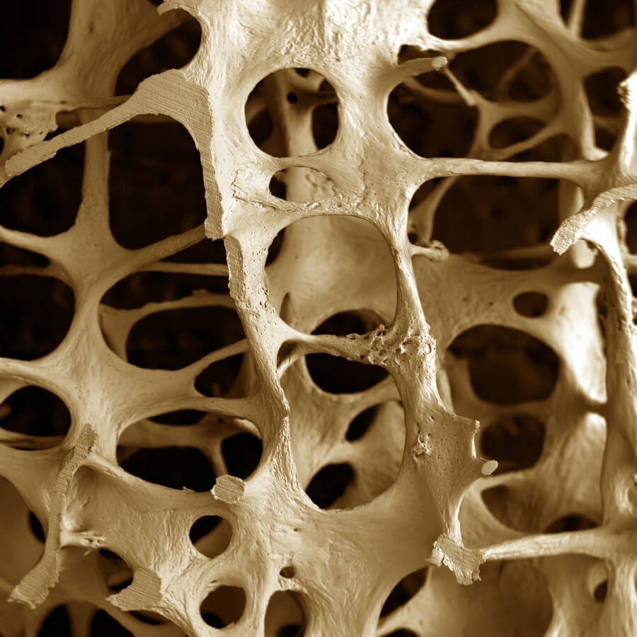
3) Spongy bone - is located under the compact substance inside the bone and is also built from lamellar bone tissue. A distinctive feature of the spongy substance is that the bone crossbars are located loosely and form cells, so the spongy substance really resembles a sponge in structure. A compact substance is found in those bones and in those parts of them that perform the functions of support and movement (for example, in the diaphysis of tubular bones). In places where, with a large volume, it is required to maintain lightness and at the same time strength, a spongy substance is formed (for example, in the epiphyses of tubular bones).
4) Inside the bone there is a medullary cavity - the walls of which are covered from the inside with a thin fibrous connective tissue sheath endosteum.
5) In the cells of the spongy substance and the bone marrow cavity there is a red bone marrow - in which hematopoiesis processes take place. In fetuses and newborns, all bones form blood, but with age, gradually, myeloid (hematopoietic) tissue is replaced by fatty tissue and the red bone marrow turns yellow and loses the function of hematopoiesis (in domestic animals, this process begins from the second month after birth).
6) Articular cartilage - covers the articular surfaces of the bone and is built from hyaline cartilage tissue.
Thus, in the bones of an adult animal, the following are isolated in layers:
periosteum, 2) compact substance, 3) spongy substance, 4) medullary cavity with endosteum, 5) bone marrow, 6) articular cartilage.
Bone classification
The following types of bones are distinguished by shape:
1) Long bones are arcuate (ribs) and tubular.
2) Short (spongy) bones.
3) Flat bones are involved in the formation of the walls of the cavities and belts of the limbs, performing a protective function (bones of the skull roof, sternum, scapula, pelvic bones).
4) Cmixed bones. Example, occipital bone.
5) Airborne bones have a cavity in their body (sinus, sinus), lined with a mucous membrane and filled with air (maxillary, frontal, sphenoid).
According to their origin, they are:
1) Primary bones.
2) Secondary bones.
The formation of bones on the basis of cartilaginous rudiments proceeds as follows.
Replacement of cartilage with bone includes perichondral and endochondral ossification.
Perichondral ossification begins with the appearance of osteoblasts from the inside of the perichondrium in the middle part of the diaphysis. The cartilage cells inside the perichondral girdle dissolve, the strength of the diaphysis increases. At this point, the perichondrium becomes the periosteum, and the perichondral ossification becomes periosteal. Blood vessels grow into the resulting cavities. Arises endochondral bone. In the future, the periosteal and endochondral bones grow in parallel. By the end of the fetal period, additional ossification points may appear in the bones - apophyses, appear where the bones have significant protrusions, tubercles. The ossified diaphysis and epiphyses are connected in tubular bones by cartilaginous plates - metaphyseal cartilages - growth zones. Due to the metaphyseal cartilage, the bone grows in length, with their ossification, bone growth stops.
The skeleton is the foundation musculoskeletal system, the main base of the organism. It consists of bones that serve as a support for all soft tissues. What is in the bones themselves, because it is impossible to imagine them empty?
Bone is an organ, and like any other, it is made up of several types of tissue. One of the main ones is a compact bone substance, without which bone formation is impossible in principle. It is adjacent to an important spongy substance. Their opposition will be discussed below.
Bones come in several types and differ from each other not only in size. Each of them has an individual purpose. In connection with the assumed bone, it occupies the most suitable location in the skeleton. The same principle applies to bones.
Therefore, compact bone tissue, more precisely, its larger amount is located in the bones responsible for the mobility of the skeleton, as well as those that perform the function of support.
The following bones do not do without a compact substance:
- Long. Responsible for the skeleton of the limbs. Their tubular middle part is completely filled with compact matter;
- Flat. Their outer part is covered with a compact substance;
- Short. Compact bone tissue also covers them from the outside, in a thin layer.
The structure of the compact substance of the bone
For a better understanding of the structure of compact bone tissue, you should first familiarize yourself with the structure of the bone as a whole.
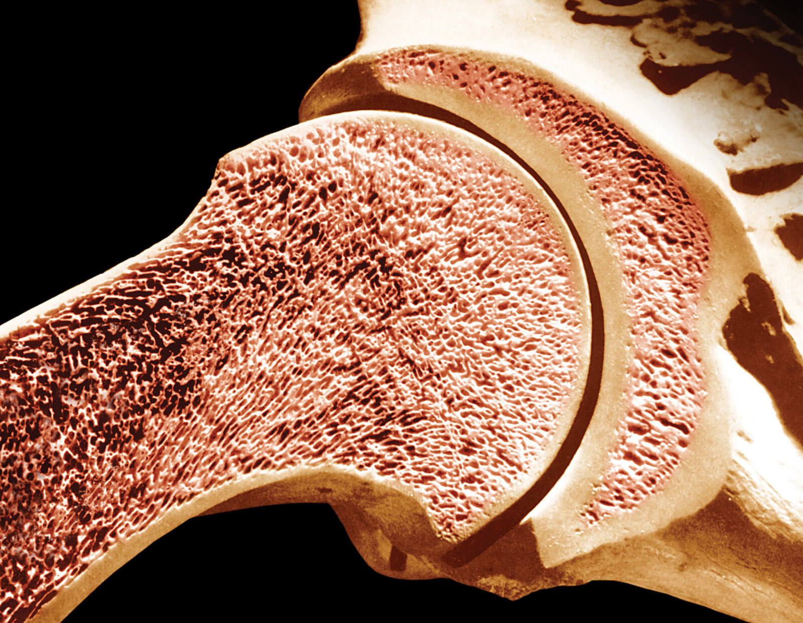
Taking a section of the bone and magnifying it with a microscope, you can see many bone plates centered around a special channel that contains nerves and blood vessels. These plates are a system called Osteon. It is the main structural unit of bone.
Such plates are ordered in accordance with the load that the bone takes on. The osteons then organize into larger bony elements called trabeculae. And only then the bone substance of two types is formed.
The whole process depends on the density of formation of these bone elements:
- In the case when trabeculae lay down in a loose plane, special cells are formed that resemble a spongy surface. This is how spongy bone tissue is formed;
- When trabeculae lie down in a dense layer, a compact bone substance is formed.
The difference between the two types of bone substance is that spongy tissue is responsible for lightness and elasticity, and therefore has a significantly reduced density. Compact bone tissue forms the entire cortical layer of bones. This is due to its high density and strength of the structure. Therefore, this substance is quite heavy and makes up the bulk of the bones of the skeleton.
Thus, the compact substance of the bone consists of the primary structural unit of the osteon, which is mainly responsible for its strength.
Learn about the structure of the skeleton from the proposed video.
Functions of compact bone tissue
In childhood, children often hear from their parents a call for active participation in sports or gymnastics. Unfortunately, not everyone follows the advice of their elders and only over time they understand how important parental phrases were.
Considering the reason for the above, one should pay attention to the following: the bone substance is divided into two types, each of which has a different composition. While the spongy substance is formed from organic chemical elements (ossein), the compact substance of the bone consists of inorganic substances. Their main composition is lime phosphate salts. They are responsible for the firmness of the fabric.
The small organism a large number of ossein, which determines the flexibility of growing bones. When the process of bone growth approaches the completion phase, some cartilage is replaced by bones, and the bones themselves acquire the necessary number of hardened protrusions and depressions on which ligaments and the muscle system are attached.
The more muscle mass the body accumulates during the period of growth, the greater the number of necessary irregularities that the bones have time to create. Then the compact bone tissue forms a dense cortical layer, and the structure of the skeleton is practically not subject to further changes.
As can be seen, the compact tissue comes into full action secondarily, after the spongy one. This is the reason for the main protective function bones.
Also, the compact substance of the bone stores everything chemical elements needed by the bones. It is it that contains in its structure a large number of nutritional holes through which blood vessels carrying nutrition penetrate.
Due to the well-coordinated work of the compact substance, nerves and vessels of the bone, it has the ability to grow in thickness, which is necessary.
The compact substance of the bone, making up most of the bone structure, forms its bulk. Fulfilling main function protection of the skeleton, and hence the support of the whole organism as a whole, a compact substance, with age, requires sufficient attention, in the form of additional sources of mineral elements, namely vitamins A, D and, of course, calcium.
Noticed an error? Select it and click Ctrl+Enter to let us know.
Mar 18, 2016 Violetta Doctor
The human skeleton is a complex of bones and their joints. It is the passive part of the musculoskeletal system, the active element of which, you guessed it, is the muscles. The mass of the skeleton on average in men is 10 kg, in women - 6-8 kg.
The human skeleton is divided into axial and accessory. The axial one is more complex, and this is understandable, because it includes such components as the skull, spinal column and chest bones. The accessory skeleton is represented by the bones of the upper and lower limbs.
The functions of the skeleton in the body are important and varied. First of all, it serves as protection for vital organs. The skull reliably protects the brain, the organs of hearing, vision, smell, the initial sections of the digestive and breathing apparatus. AT spinal canal contains the spinal cord. Rib cage serves as protection for the heart, lungs, thymus gland, esophagus and large vessels. In the pelvic cavity are bladder, as well as the uterus, vagina, tubes, ovaries in women and the prostate gland in men.
The skeleton is also a support for soft tissues and organs. It determines the external form of individual parts of the body and the entire human body as a whole. Movement is provided by bones that are movably connected to each other, set in motion by muscles.
And of course, we are interested in the biological function of the skeleton, namely its participation in mineral metabolism. Although the biological function of the skeleton also includes hematopoiesis and immunity.
Now let's talk about the bone as an organ. Perhaps, for some, such a combination of the words “a bone is an organ” is not quite familiar. Nevertheless, this is true: the bone is the same organ of the human body as all the rest. Each of the more than 200 bones of the skeleton is a living, actively functioning and continuously renewing organ. Vessels and nerves penetrate into the bone, as well as into all other organs, providing nutrition to the bone tissue and its interaction with the whole organism.
Each bone has its own development and shape, occupies its own place in the body, always connects with other bones (except for the hyoid bone and sesamoid bones, located in soft tissues). Each bone consists of representatives of all 4 types of tissues: connective tissue, endothelium, muscle and nervous tissue. All together they form a bone structure that can be rebuilt very quickly under the influence of external and internal factors. Let's remember this recipe for bone health, so to speak, that point, that feature of bone tissue (which you may not have suspected), which allows you to influence the vital activity of the bone, its metabolism consciously. This is very nice, and we will definitely use it in the chapter on home workouts. In the meantime, we continue our excursion into the science of osteology!
The main ones in the bone, of course, are the bone cells. The functional element of the bone are special cells - osteoblasts. These cells are able to produce a special protein substance for bone - ossein, as well as deposit mineral salts. Osteoblasts are located in the inner layer of the periosteum and are involved in the growth of the bone in width and the restoration of its integrity after fractures.
The bone is actively involved in metabolism, is constantly under the influence of nervous system, hormones, nutritional conditions of the body, the degree of physical activity. I will always draw your attention to the fact that physical exercise for bones are necessary. I hope you will remember this very soon and begin to feed your bones as much as possible. Now you understand that bones, like all other organs, make up a very dynamic system.
On external examination, the bone has a yellow color, the ends are covered with blue-white cartilage. Outside, every bone except articular surfaces, has a periosteum, i.e., a connective tissue sheath.
The difference in the conditions under which bone develops internal structure and the functions performed - all this determines the diversity of bone shapes.
Tubular bones, long and short, they distinguish an elongated cylindrical part, called the body, or diaphysis. At each end of the body (diaphysis) is the epiphysis. There are two epiphyses, respectively. On a cut (cut) in the area of the diaphysis, a cavity is visible, which in adults is filled with yellow bone marrow. In fetuses and newborns, the bone cavity is absent, and there is red bone marrow in the diaphysis.
The wall is formed by the hard substance of the bone. The epiphyseal ends are more massive than the diaphysis, and are formed by a spongy substance, in the cells of which there is a red bone marrow. Tubular bones mainly make up the skeleton of the limbs, providing extensive movements.
Spongy bones are covered on the outside with a thin plate of solid substance, and inside they are filled with plates of spongy substance. They do not have a medullary cavity, like tubular bones. The red bone marrow is located in small spongy cells separated by bone beams oriented in the direction of the force acting on this bone.
Fractures in osteoporosis occur in places where spongy tissue is located, and these are the end parts of tubular bones, vertebrae, small bones of the wrist and pelvic bone. spongy bone especially susceptible to osteoporosis.
Flat bones have well-developed compact outer plates, and between them there is an insignificant layer of spongy substance.
Pneumatized (air-bearing) bones have sinuses that communicate with the nasal cavity, and mastoid cells communicate with the tympanic cavity.
The flat bones of the skull, spine, sternum, shoulder blades, ribs, and pelvis contain the bone marrow, which carries out hematopoietic and immune functions. The bone is involved in the exchange - when necessary, the body sucks out minerals from it (most often during stress), and then does not always give it back. The bones of the skull work like pumps, distributing CSF throughout the skull and spinal canal. Bones have different properties: in the ethmoid and frontal bones there are labyrinths with which the air is warmed. Bones, especially labyrinths temporal bones, can be resonators, helping to receive a danger signal.
There are 3 types of cells in bone: osteoblasts, osteocytes, and osteoclasts.
osteoblasts(we have already mentioned them) - young bone cells. They have high energy capabilities, can secrete many different enzymes and are located in the form of beams at ossification points in the surface layers of the bone. Gradually, the beams grow in all directions, forming a mesh network, the cells of which contain blood vessels and bone marrow cells. Osteoblasts produce proteins and intercellular substance, which is then impregnated with calcium salts. So they themselves are immured in the bone substance and turn into osteocytes.
Osteocyte- Mature bone cell. Osteocytes are located in the cells of the bone network, surrounded by tissue fluid, due to which they are nourished and cleaned. osteoclasts- large multinucleated cells. Osteoclasts break down bones and cartilage in the process of bone renewal. They have numerous outgrowths, and this increases the area of contact between osteoclasts and bone.
The outer layer of the bone is a compact substance that looks like a dense, but on the cut of a shiny plate. The bodies of tubular bones are built from a compact substance. The basis of the compact substance is an intermediate substance in which osteons are located - the structural units of the bone. What it is? Osteon is from 4 to 20 tubes of intermediate substance inserted one into the other. In the center of the osteon there is a channel with a diameter of 10-110 microns, through which a blood capillary passes. With their length, the osteons are oriented perpendicular to the plane of pressure. Osteons do not come into contact with each other, between them there are intercalated plates, which unite the osteons into a single whole.
Each bone contains a huge number of osteons. There are about 3200 of them in the femur. If we assume that on average each osteon consists of 12 tubes, then there will be 384,000 of them in the femoral shaft, inserted one into the other. Therefore, with such an architecture, the femur can withstand a load of 750 to 2500 kg.
Features of the structure of the bone with a relatively small cost of material provide its greatest strength. The number, thickness and shape (round, oval, irregular) of the osteon tubes can change under the influence of muscle work, pressure and stretching forces, or other factors associated with the profession, nutritional conditions, and metabolism. The restructuring of osteons will also affect the strength of the bones. It should be clear what causes such a margin of bone tissue strength: bones sometimes experience quite large loads, for example, when jumping from a run or from a height.
The spongy substance is located under the compact and is built from bone thin crossbars, with their edges located perpendicular to the lines of compression and tension. These crossbeams form columns with each other, intersecting at an angle of 90°, and at an angle of 45° intersect the long axis of the bone. The crossbeams are oriented at one end in the direction of pressure forces, and at the other they rest on the compact substance of the bone. As a result of this, the forces are decomposed into two components, which are the sides of the parallelogram of the force, along the diagonal of which the force is distributed evenly to the walls tubular bone from any articular surface.
Most bulk part bone is an intermediate (basic) substance representing the product of osteoblasts.
There are a lot of osteoblasts in the growing bone, especially under the periosteum and in the area of the epiphyseal cartilage. In an adult, when bone growth is completed, these cells are found only in areas of bone tissue restoration (with fractures and cracks in bones). Thus, in each bone in different age periods there is a certain quantitative combination of cellular elements: osteoblasts, osteocytes and osteoclasts, which create new bone substance, destroy the old one and ensure the stability of bone metabolism.
The intermediate substance consists of collagen fibers (organic) and mineral salts (inorganic), which impregnate bundles of collagen fibers. The combination of organic and inorganic substances creates an elastic and solid structure.
On the example of the structure of bone tissue, the relationship between structure and function is clearly visible. This is especially easy to notice when the function of movement is disturbed or changed. In this case, a significant restructuring of the architecture of compact and spongy matter occurs. With a decrease in the load on the bone, part of the bone plates atrophy and architecturally rebuild, and, conversely, an increase in the load on the bone has a formative effect.
Well, thin women, now it’s clear why you are shown athletic gymnastics? Bones do not have enough weight to be strong. There is such a term in medicine - "the risk of developing a disease." With osteoporosis, there is a long list of things that make it more likely. If possible, we will consider how exactly this or that factor can cause the onset of osteoporosis, so that you can then decide for yourself how important all this is for you. A conscious approach is possible when there is an understanding of the essence, and we now need just such an approach.
Periosteum - the outer surface of the bone (with the exception of the articular surfaces and places of attachment of the tendons), is a thin (100-200 microns) plate. The periosteum is tightly attached to the bone due to the presence of special fibers penetrating perpendicularly into the compact bone substance. The periosteum consists of two layers - outer and inner. There are many collagen fibers in the outer layer, among them are nerves, plexuses of small arteries, veins, and lymphatic vessels. Blood vessels give the periosteum a pink hue. The fibrous layer of the periosteum is adjacent to the bone and contains osteoblasts, which, as the bone grows in thickness, form the common (general) outer plates of the intermediate substance.
The composition of the living bone of an adult includes water 50%, fat 15.75%, ossein (collagen fibers) 12.4%, inorganic substances 21.85%. Dried bone is 1/3 organic and 2/3 inorganic. Inorganic substances are various salts (lime phosphate - 60%, lime carbonate - 5.9%, magnesium sulfate - 1.4%). In addition, there are various chemical elements in the bones. Mineral salts dissolve easily in a weak solution of hydrochloric or nitric acid. This process is called decalcification. After such treatment, only organic matter remains in the bones, which retains the shape of the bone. It is porous and elastic, like a sponge. When organic matter is removed by burning, the bone also retains its original shape, but becomes brittle and crumbles easily. Only a combination of organic and inorganic substances makes the bone hard and elastic. Its strength is greatly enhanced by the complex architecture of the compact and spongy substance.
Bones have plasticity, are easily rebuilt under the influence of training (moderate and regular is best), which is manifested in a change in the number of osteons and the thickness of bone plates. Bone remodeling occurs due to the formation of new bone cells and intercellular substance against the background of bone destruction by osteoclasts. Lack of load leads to weakening and thinning of the bone. The bone becomes coarse and partially resorbed - this is osteoporosis.
And now let's briefly repeat the technology of bone tissue reconstruction. Osteoclasts destroy bone, they do it at the request of the body, when it needs additional calcium. Osteoclasts secrete a special substance (acid), which dissolves the old bone. As a result of this dissolution, many minerals enter the blood, including calcium.
As you understand, the result of such work is a cavity. You can’t leave it like that, and the command for repair goes to other cells (I think you have already guessed which ones) - osteoblasts. Osteoblasts first line the resulting cavity with collagen - a viscous adhesive substance (as they cover with glue), and then draw calcium and other trace elements from the blood, forming crystals on the surface of the "glue". All this gradually hardens, turning into bone. And osteoblasts after such work cease to be osteoblasts, they lose their activity, immured in the bones and from that moment are called mature cells - osteocytes. The entire reconstruction cycle takes from 3 to 6 months, frankly, it does not happen quickly.
If for various reasons osteoclasts are more active than osteoblasts, then bone resorption is incomparably faster than its restoration. This is how bone is lost. I would like to know what can change the activity of cells in the direction of bone destruction. This, in fact, is the answer to the question, for what reason this unnecessary mechanism is launched for the occurrence of osteoporosis. Let's figure it out.
Many factors are involved in the process of bone tissue reconstruction. The first is the endocrine system. Parathyroid hormone - parathyroid hormone increases bone destruction by activating osteoclasts. The hormone calcitonin, which is produced in thyroid gland and is opposite in action to parathyroid, enhances the processes of bone formation, stimulating the activity of osteoblasts. thyroxine, a hormone thyroid gland, and cortisol, the main hormone of the adrenal glands, enhance the processes of bone tissue destruction. Vitamin D, which is involved in the regulation of calcium absorption in the intestine, plays a certain role in calcium metabolism and, consequently, in the development of osteoporosis.
What is the role of female sex hormones in this? And this noble role is protective, and it is realized as follows.
1. Female sex hormones are able to suppress the activity of parathyroid hormone.
2. Estrogens are able to suppress the destructive effect of thyroxin on bone tissue by increasing the synthesis of thyroxin-binding protein, i.e., female sex hormones act on thyroxin indirectly, through a special protein that is able to bind thyroxin and thereby make it inactive.
3. Osteoblasts have estrogen-sensitive receptors. This means that female sex hormones have the ability to directly affect osteoblasts, and osteoblasts become more numerous.
4. Estrogens increase the return of calcium to bone tissue.
Along with the opinion of official medicine, I am pleased to offer you a version of osteoporosis by a healer from Novosibirsk, I. A. Vasilyeva.
There is a connection between bone and endocrine glands. The bone is destroyed when the defenders are weakened, injuries, stress (high levels of cortisol and parathyroid hormone).
The main causes of bone destruction are:
1) injuries of the skull, pelvis and spine;
2) post-traumatic scoliosis of the spine;
3) foci of osteoporosis that have arisen near the site of injury;
4) An increase in the level of parathyroid hormone also leads to a decrease in calcium and magnesium ions in the blood serum;
5,) malnutrition of the cervical sympathetic nodes, thyroid and parathyroid glands (due to cervical scoliosis);
6) weakening of pancreatic function and a drop in insulin levels;
7) inflammatory foci in the region of the skull;
8) venous congestion in the veins of the intestine (pelvic bones suffer from injury), liver (suffer lumbar spine);
9) long pathological conditions with low blood volume.
The biggest enemy of the bone is trauma. Injury impairs the blood flow of the bone itself: inflammatory foci appear in the bone and adjacent tissues, and this already disrupts the operation of the control system and blood supply to the body as a whole. Then the bone not only does not have enough blood, it is prevented by near stagnation of blood, and the bone does not receive what it should receive. Then the bone loses its function and changes its structure.
The bottom line is that it is the border tissues - bones and epithelium - that get the bulk of injuries (breakdowns). And it is the bones and epithelium, to a greater extent than other tissues, that are characterized by unconscious regulation. This reaction of the connective tissue is the greatest danger to the body.
How does the process of reducing bone mineral density occur?
Calcium is washed out of the bone into the space surrounding the bone. Organs that need calcium, functional systems or foci (pseudo-organs), and secrete the appropriate enzymes. The mineral density of bone tissue is reduced in the bones at the site of injury near inflammatory foci. Mineral density is reduced because inflammatory foci contribute to the "washout" of calcium from the bone. In this case, the spent calcium is ejected directly into the intercellular substance. The concentration of calcium in the lymph increases, kidney and gallstones, tubules and capillaries overgrow on the bones. Spondylarthrosis develops (narrowing of the intervertebral foramina) and compression of the nerve roots, followed by the development of nervous disorders.
The skeleton is, among other things, also a depot of calcium. When everything is in order in the body, calcium is used sparingly. But, it turns out, it happens differently.
| |