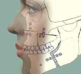
The upper jaw is a paired bone that is located in the center of the front of the face and connects to the rest of its bones.
Performs a number of important functions: participates in the functioning of the masticatory apparatus, in the formation of cavities for the nose and mouth, and the partitions between them.
Anatomy upper jaw The human body has a complex structure. It consists of a body and 4 processes - alveolar, where the cells of the teeth are located, frontal (directed upward), palatine and zygomatic.
The upper jaw is much thinner, and it is also quite light due to the sinus (cavity), with a volume of approximately 4-6 cm3.
The body of the jaw consists of the anterior, infratemporal, nasal and orbital surfaces. The front includes a hole where thin blood vessels and nerve processes.
Blood supply occurs through 4 alveolar openings in the infratemporal area.
The nasal surface forms turbinate, and the flat orbital contains the lacrimal notch.
The upper jaw is motionless due to fusion with the bones of the face, has almost no connection points masticatory muscles and is under the influence of pressure rather than traction.
Frontal process
(lat. processus frontalis)
The frontal process of the maxilla is directed upward and connects to the nasal part of the frontal bone. It has a medial and lateral zone. The medial region of the frontal process includes the lacrimal crest. The posterior part borders the lacrimal groove.
Palatine process
(lat. processus palatinus)
The palatine process of the maxilla is part of the system of hard tissues of the palate. It has a connection in the form of a median suture with the process of the opposite side, as well as with the plates of bones. The nasal ridge is formed along this suture. The palatine process has a smooth surface above and rough below.
Alveolar process
(lat. processus alveolaris)
Alveolar process The upper jaw consists of an outer (buccal), inner (lingual) wall, as well as dental alveoli made of spongy substance where the teeth are placed. The complex structure of the alveolar process also includes bone partitions (interdental and interradicular).
Anterior surface of the body
(lat. fades anterior)
The anterior surface of the body borders the infraorbital margin. It has a hole with a diameter of 2-6 mm, under which there is a fang pit. There begins the muscle that is responsible for raising the corner of the mouth. The front surface of the body has a slightly curved shape.
Infraorbital foramen
(lat. foramen infraorbitale)
The infraorbital foramen is located on the anterior surface of the body approximately at the level of the 5th or 6th tooth. The thinnest blood vessels pass through it, as well as processes trigeminal nerve. The diameter of the infraorbital foramen is quite large (can reach 6 mm).
Zygomatic process
(lat. zygomaticus)
The zygomatic process of the maxilla starts from the upper outer corner of the body. It is directed laterally (relates to the side of the surface) and has a rough end. The zygomatic process of the frontal bone connects with the temporal process.
Posterior (infratemporal) surface of the body
(lat. facies infratemporalis)
The posterior surface of the body is separated from the anterior surface with the help of the zygomatic process and has an uneven, often convex shape. Here is the tubercle of the upper jaw, where the alveolar canals open. On the side of the tubercle of the posterior surface of the body there is also a large palatine groove.
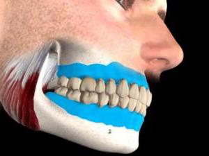
This article is aimed at conveying to the reader information about the general structure of the upper and lower jaws of a person; special attention will also be paid to the alveolar processes, an important component of our masticatory and communicative apparatus.
Delving into the upper jaw (HF)
The maxillary part of the human cranial bones is paired. Its location is the central front part. It fuses with other facial bones, and also articulates with the frontal, ethmoid and sphenoid. The upper jaw is involved in the creation of the orbital walls, as well as the oral and nasal cavities, the infratemporal and pterygopalatine fossae.
In the structure of the upper jaw there are 4 multidirectional processes:
- frontal, going upward;
- alveolar, looking down;
- palatal, medially facing;
- zygomatic, laterally directed.
The weight of the human upper jaw is quite small, it does not seem so upon visual inspection, and this is due to the presence of cavities, for example the sinus (sinus maxillaris).
A number of surfaces are also distinguished in the structure of the upper jaw:
- front;
- infratemporal;
- nasal;
- orbital.
The anterior surface originates from the level of the infraorbital margin. Just below there is a hole along which nerve fibers and blood vessels run. Below the opening is the pterygopalatine fossa, in which the beginning of the muscle responsible for raising the oral corners is fixed.
The surfaces of the orbits are covered with lacrimal notches. On their areas remote from the anterior edge there are grooves, one on each, called infraorbital.
Most of the nasal surface is occupied by the maxillary cleft.
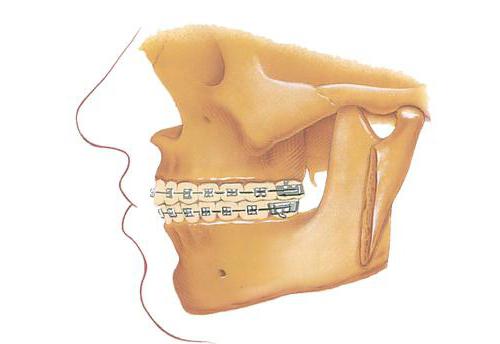
Alveolar component
The alveolar process of the maxilla is part of the maxillary body of the bone. It is united by an intermaxillary suture with the outgrowths of the jaw located on the opposite side. Without a visible feature from behind, it changes, turning into a tubercle facing the process of the palate of the upper part of the jaw. At the same time, he looks medially. Its shape is similar to an arc that is curved like a bone ridge, which has a forward-facing convexity.
The outer surface turns into the vestibule of the mouth. It is called vestibular. The inner surface faces the sky. It's called palatal. The alveolar process on its arch has 8 alveoli of different size and shape, intended for molars. The alveoli of the incisors and canines include two main walls, labial and lingual. There are also lingual and buccal walls. But they are located in the premolar and molar alveoli.
Functional purpose
The alveolar processes have interalveolar septa made of bone tissue. Alveoli, which are multi-rooted, contain septa that separate the roots of the teeth. Their size is similar to the shape and size of tooth roots. The first and second alveoli include incisal roots, which look like cones. The third, fourth and fifth alveoli are the location of the roots of the canines and premolars. The first premolar is often divided by a septum into two chambers: buccal and lingual. The last three alveoli contain the roots of the molars. They are separated by an interroot partition into 3 root compartments. Two of them address the vestibular surface, and one - the palatine surface.
The anatomy of the alveolar process of the upper jaw is designed in such a way that it is somewhat compressed on the sides. As a result, its size, like the size of any of these processes, is smaller in the direction from front to back than in the buccal-palatal region. The lingual alveoli have a rounded shape. The variable number and shape of the dental roots of the third molar cause it different shapes. Behind the 3rd molar there are plates, external and internal, which, converging, form a tubercle.
Features of the parameters of the upper jaw
The individual shapes of the upper jaw in people vary, as do the shapes of its alveolar processes. However, in the structure of the jaw, two extreme forms can be distinguished:
- The first is characterized by narrowness and is itself tall.
- The second is wide and low.
The shapes of the pits of the alveolar processes, accordingly, may also differ slightly depending on the type of jaw structure.
On this jaw there is a maxillary sinus, which is considered the largest of the paranasal sinuses. Its shape is usually determined by the shape of the maxillary body.
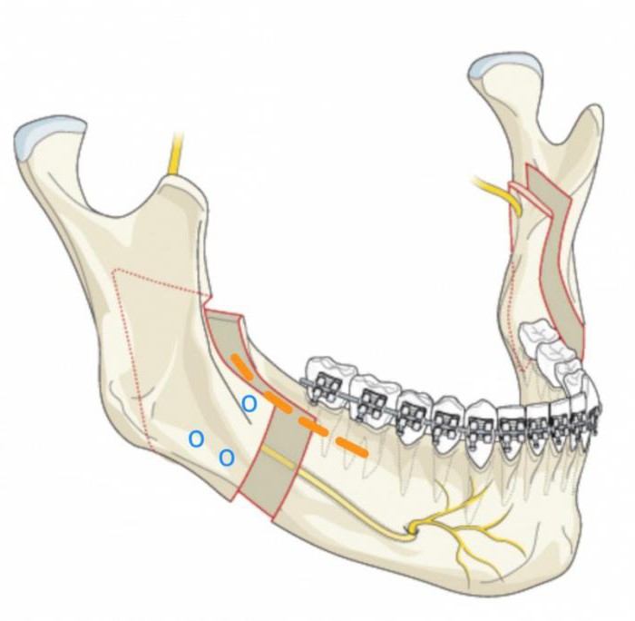
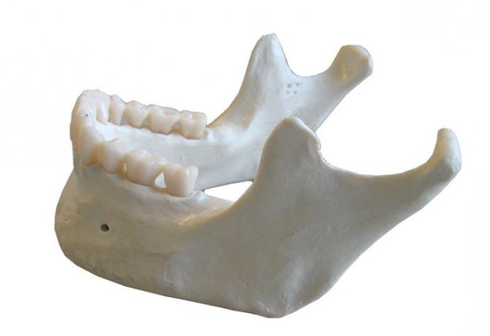
General data about the lower jaw (LM)
Bone lower jaw takes its development from two arches: the gill and the first cartilaginous. The size of the lower jaw is significantly smaller than that of human predecessors, which is due to the emergence of oral speech in humans. And also large sizes the lower jaw would interfere to modern man when chewing food, due to its location when planting the head.
In the lower jaw there are such structural elements, How:
- alveolar process - the outermost part of the jaw body in which the dental cells are located;
- mandibular body;
- chin hole;
- mandibular canal;
- mandibular angle;
- branches of the jaw;
- a number of articular and coronoid processes;
- opening of the lower jaw;
- head.
The resulting shoots
The bone in question has the alveolar process of the mandible. The alveolar composite contains eight dental sockets on both sides. These alveoli are separated by septa (septa interalveolaria), and their walls face the lips and cheeks. They are called vestibular. The walls face the tongue. On the surfaces of the alveolar bodies, a raised formation (juga alveolaria) can be clearly seen. In the place between the protrusion of the chin and the alveolar incisors there is a sub-incisal depression.
The depth and shape of the alveolar process can be varied, in accordance with the shape and structure of NP formation. The alveoli belonging to the canines are round in shape, and the deep alveoli belong to the second premolar. Each molar has bony septa between the root attachment sites. The alveolus of the third molar can vary among individuals in appearance and the presence of the number of septa.
In the LF, the alveolar process has a similar structure to the alveoli of the HF. They have two-thirds walls: lower and upper. The upper third is formed by plates of hard and compact substance, and the lower third is lined with spongy-type tissues.
Summing up
Now, having general information about the structural components of the upper and lower jaw, knowing their location and function, you can characterize them. In addition, the structure of the alveolar processes of these jaws, the presence of special components in them and their functional purpose were examined. We also saw that the alveoli of both jaws are largely similar to each other and can slightly change their shape depending on the type of jaw structure.