Respiratory system
In reptiles, the lungs are the only respiratory organ. The respiratory tract begins with the nasal cavity, which has nostrils (external respiratory openings) and choanas (internal respiratory openings), which open further into the oral cavity. Then comes the larynx and trachea Behind the trachea branches into 2 bronchi (tubes). Each bronchus enters a corresponding lung. The lungs of lizards and snakes have a fine-meshed structure; crocodiles and turtles also have internal partitions that increase the area of gas exchange. Air is sucked into the lungs, and then thrown back due to the contraction of the intercostal muscles, which change the volume of the chest. At the same time, the lungs are also stretched, and air is sucked in through the respiratory openings. Only turtles have lost the ability to breathe like this. In them, the filling of the lungs occurs, as in amphibians, by swallowing air.
Like other reptiles, lizards have a single occlusal condyle. Ribs are present on all vertebrae except the caudal and cervical vertebrae. Several families of lizards may voluntarily shed their tails captured by a predator. It is believed that the writhing tail, which is thrown back, distracts the attention of the predator, allowing the lizard to escape.
Structure and life
In a clinical scenario, autotomy may occur when the lizard is held. It is important to be prepared for this event. Lizards capable of autotomy have a vertical plane of destruction through the body and part of the neural arch of the caudal vertebrae. This is a plate of cartilage or connective tissue that develops after ossification. Spasms occur in the vasculature supplying the tail when autotomy occurs, which stops any bleeding.
Support- propulsion system
The skeleton of a reptile consists mainly of bone tissue, which increased its strength, so the size of reptiles is larger than that of amphibians. The skeleton consists of 3 sections - the skull (skeleton of the head), the spine (skeleton of the body), the skeleton of the limbs and their belts.
The skeleton of the head (skull) is all bony, has an elongated shape (due to elongation of the bones). The volume of the cerebral part of the skull increases. A hard palate appears, which separates the nasal and oral cavities. The skull is connected to the spine with the help of one condyle of the occipital bone. Atlas and epistropheus (the first 2 cervical vertebrae) form a movable joint with the skull. The mobility of the head is also provided by a flexible neck. Increased head mobility helps animals better navigate their environment.
The spine is made up of five sections:
- cervical (8-10 vertebrae);
- thoracic region (5 vertebrae);
- lumbar (17 vertebrae);
- sacral (2 vertebrae),
- tail section (several tens of vertebrae).
In the trunk part of the spine in reptiles is divided into 2 sections - the thoracic and lumbar. The ribs are attached to the thoracic vertebrae. The spine in snakes is formed by only 2 sections - the trunk section and the tail. The skeleton of the limbs and girdles of the limbs, as well as the sternum, are reduced in snakes.
The limbs of reptiles, by the nature of their structure, occupy an intermediate place between amphibious vertebrates and mammals. The limbs and limb belts of reptiles are firmly strengthened. In the forelimb, the shoulder and forearm are lengthened, and in the hind limb, the thigh and lower leg are lengthened. But the size of the final sections, on the contrary, decreases (feet and hands). Separate parts of the limbs change their position in relation to each other and the body. The unequal arrangement of the departments in the limbs corresponds to different functions: the forelimb, when moving the reptile, pulls the body, the hind limb pushes it. In higher terrestrial vertebrates, differences in the position of the limbs extend to the thigh and shoulder, and in lower ones, to the lower leg and forearm. The ribs of reptiles are fused with the sternum, and this greatly strengthens the belt of their front limbs, which, due to this, are connected with the spine. The pelvic girdle is a strong support for the hind limbs. Reptiles have claws on their fingers.
The muscles of reptiles are more differentiated than those of amphibians. The muscles of the neck, abdominals, intercostal muscles and subcutaneous muscles appear. Both flexors and extensors of the fingers appear.
If the tail is completely dropped, it cannot be retracted due to loss of blood supply to the tail. The animal is able to grow a new tail - growth usually starts after about one month and ends after about one to two years. The regenerated tail is never as long or well formed as the original, and may have a slightly different scale and pattern.
§34. musculoskeletal system of vertebrates
Respiratory diseases in captive reptiles are all too common, unfortunately, and are a great source of frustration for owners and veterinarians, as well as serious animal care for affected animals. There are many causes, including parasitic, bacterial, viral, and fungal infections, as well as traumatic injuries, tumors, and heart disease. Respiratory diseases in reptiles are often more severe and difficult to treat than in mammals due to the differences in anatomy and physiology described below.
§34. MUSCLE-MOTOR SYSTEM OF VERTEBRATE ANIMALS
WHAT TYPE OF THE LOCOMOTOR SYSTEM IS CHARACTERISTIC FOR CHORD ANIMALS?
All chordates have an internal skeleton that performs protective function and serves as an attachment site for muscles. During embryonic development, the notochord is laid in these animals. In vertebrates, the notochord is replaced by the cartilaginous ( cartilaginous fish, sturgeons) or bone (bone fish, amphibians, reptiles, birds, mammals) spine. The spine is built from vertebrae (Fig. 201). Each vertebra consists of a body and an arch. The holes of the upper arcs, overlapping each other, form spinal canal in which the spinal cord is located.
Hopefully this will give you a better idea of how these conditions can be prevented, what to look for when they occur, how they can be treated, and why they can be so difficult to treat. I had my work and reputation criticized by colleagues and no doubt in the forums when "the antibiotics he dispensed didn't work."
These were invariably cases where the owner did not follow all the other suggestions that come with the medications, when they brought the animal to me in a state of illness and were informed of the guarded prognosis, but decided to try the treatment anyway, where they did not return for me. to understand why my treatment of choice didn't work. Enough with the buy-trilbashi! Successful results rely on a holistic, integrated approach as well as the hobbyist who works with the veterinarian, not against.
The skeleton of a bony fish consists of a skull, spine, and fin skeleton. Skull - the skeleton of the head, formed by a large number of bones that are fixedly interconnected (Fig. 202. A). Only the lower jaw is movably connected to the skull, which enables the animal to actively capture prey and gill covers. The skull protects the brain. The structure of the skull also includes gill arches, on which the gills are located. In the spine of fish there are two given away: tulubovy and caudal. The tubular vertebrae bear ribs, but do not have caudal ribs.
The difficulty in treating respiratory problems in reptilians is primarily due to their inability to effectively clear fluid, debris, and waste from their respiratory tract by coughing. Most species other than crocodilians do not have a true diaphragm and therefore do not possess the cough reflex. The mucociliary apparatus is poorly developed in the airways of reptiles, making it difficult to clear infectious agents and subsequent secretions. This is a vital defense against respiratory infection and disease that is lacking in reptiles compared to mammalian species.
The fin skeleton of bony fish is represented by bony rays. The skeleton of paired fins (pectoral and ventral) also includes limb belts. Attached to their bones are muscles that move their fins. The muscles are better developed on the dorsal side of the body and in the tail section (Fig. 202. B). The muscles of the body look like wide ribbons. There are also specialized muscles that move lower jaw, gill covers, fins and the like.
In this way, secretions and infectious material are combined in the lower parts of the lungs, which makes penetration by immune cells as well as drugs very difficult, resulting in chronic diseases which are often difficult to treat. Thus, having the same expectations when treating a respiratory infection in a snake or lizard, for example, compared to a dog or cat, can lead to disappointment. The anatomy of the reptile airways is also unique in several ways that can influence disease progression.
Rice. 201. The structure of the vertebra of a mammal: 1 - body; 2 - arc; 3 - spinal canal
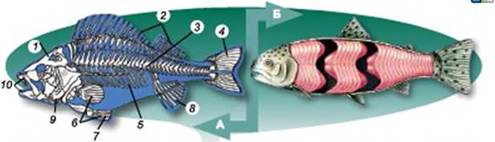
Rice. 202. A. Skeleton of bone fish: 1 - skull; 2 - skeleton of dorsal fins; 3 - spine; 4 - skeleton of the caudal fin; 5 - ribs; 6 - skeleton of pectoral swimmer; 7 - skeleton of the ventral fin; 8 - skeleton of the anal fin; 9 - gill cover; 10 - jaws (upper and lower). Would. Fish muscles (note that they are poorly differentiated)
The main anatomical value of significance in reptiles is in snakes, most of which have only one right lung. Lizards and Chekhonians have two symmetrical lungs. Boas and Pythons have a basic single right lung and a greatly reduced or rudimentary left lung. Some snakes also have a "tracheal lung", which is a disruption of the trachea with a respiratory mucosa for gas exchange.
Because of the only lung, snakes with respiratory infections are even more complex clinical cases than Chelonians and lizards. Poor farming is the single most important factor predisposing reptile-resistant individuals to disease, respiratory or otherwise. Needless to say, the specific and precise needs of individual species must be investigated and accurately assessed in order to maintain health and prevent health problems. Therefore, it is imperative that all reptiles are provided with ideal farming conditions that mimic their natural conditions and requirements in the wild as closely as possible.
The movements of fish are primarily provided by fins: the caudal fin is an active forward movement, and paired (pectoral and abdominal) - slow movement, stops and turns. Unpaired fins provide stability to the body while swimming. The spine also participates in the movement: the muscles bend the body, and the spine, when they are relaxed, unbends it.
If any issues related to raising farm animals are optimal, the animal can succumb to opportunistic infection by gradually weakening and weakening the immune system. Nutritional deficiencies are a common factor leading to such weakness and subsequent predisposition to infection.
Similarly, ascaris worms often cause damage to lung and liver tissue as they pass through the body as migratory larvae. Many pathogenic bacteria have been isolated and implicated in lung disease in reptiles, some of which are primary pathogens and others are secondary invaders causing opportunistic infections. The presence of these microorganisms does not necessarily mean that disease is present, as they may only be opportunistic to cause disease when the animal's immune system is weakened or malfunctioning.
WHAT ARE THE FEATURES OF THE MUSCULOCUTE SYSTEM OF TERRESTRIAL VERTEBRATES?
The amphibian skeleton (Fig. 203) has a cervical, tulubovy, sacral and tail sections. The only cervical vertebra that connects the head to the body. Therefore, the mobility of the head in these animals is limited. The bones of the pelvic girdle are attached to a single sacral vertebra. Amphibians usually do not have ribs, only in newts, salamanders they are underdeveloped. The amphibian skull is mostly cartilaginous.
Other pathogens may pass into the respiratory tract from other sites, primarily from generalized systemic infection or septicemia, or from infections oral cavity. Bacterial infections may be associated with viral infections, as well as with mycoplasma or mycobacteria present in the heparovirus infection that causes the chronic syndrome. The most common infections are the result of exposure to saprophytic or soil fungi in the external environment, and most are secondary pathogens to bacterial disease or traumatic injury.
The limb skeleton includes bones free limbs(front and back) and their belts. It has a structure typical of terrestrial vertebrates. The forelimb is formed by the bones of the shoulder, forearm and hand. Forelimb belt ( shoulder girdle) consists of paired bones: shoulder blades, crow bones and clavicles. The sternum joins at the junction of the crow bones. The hind limb consists of the bones of the thigh, lower leg and foot. The belt of the hind limbs is formed by three pairs of bones fused together - the bones of the pelvis. Limbs end with fingers (mainly five).
If the cornfield is inappropriate, especially with regard to ventilation and humidity, the risk fungal disease often rises. This virus is highly contagious, causing hemorrhagic pneumonia and viremia to other organ systems. It is shed in the respiratory tract and can be easily spread through a collection of poor hygiene and sanitation practices. Animals may be clinically symptom-free for extended periods with an incubation phase of up to 8 weeks, so testing during this period may be false. negative results for this reason.
The musculature of amphibians is better differentiated than that of fish. This is due to the exit to land and the appearance of paired limbs. Amphibians have the best hanging muscles of the limbs and their belts.
Thanks to paired limbs, tailed amphibians can crawl from one place to another, arching their body like a worm (give examples of tailed amphibians). In tailless amphibians, well-developed hind limbs provide jumping movement (give examples of tailless amphibians). Between the fingers of the hind limbs of frogs there are membranes, thanks to which these animals can swim.
Testing is usually based on autopsy samples, but serum testing may be performed. Another paramyxovirus associated with the parainfluenza 2 virus is involved in respiratory illnesses as well. Inclusion of the disease The body is a viral infection, mainly from boas and pythons, which can cause respiratory disease in the form of pneumonia, in addition to the more common signs of central nervous system such as "telescopes" and gastrointestinal diseases causing regurgitation and anorexia.
Thought to be a retrovirus scientists only recently discovered that the infection is actually caused by a new virus belonging to the arenavirus group. Herpesvirus is a common cause of respiratory illness in chelonians, especially Mediterranean tortoises, often with devastating consequences. Such infections often cause secondary bacterial and mixed infections. viral infections. Clinical signs include lethargy, rhinitis or "runny nose", anorexia, conjunctivitis, and stomatitis.
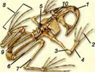
Rice. 203. Amphibian skeleton: 1 - skull; 2 - brush; 3 - shoulder; 4 - forearm; 5 - spine; 6 - thigh; 7 - lower leg; 8 - foot; 9 - tail bone; 10 - scapula
The spine of reptiles (Fig. 204) consists of five sections: cervical, thoracic, lumbar, sacral and caudal. cervical formed by several vertebrae, which provide significant mobility of the head. The thoracic and lumbar vertebrae are joined by well-hung ribs. reptiles have a real rib cage formed by the thoracic vertebrae, ribs and sternum(find it in the figure. 204). The skull of a reptile is almost entirely bony.
Finally, iridovirus infections with ranavirus, particularly from amphibian hosts, can cause severe necrotizing tracheitis and pneumonia. It is an extremely aggressive infection that causes extensive and painful tissue damage. Infections are extremely difficult to treat.
Helonian shell injury can lead to non-communicable lung diseases, often associated with dog or wildlife seizures, car accidents, and even lawnmower injuries! Conch repair techniques are tailored to individual injuries, but treating strokes and secondary infections is vital to a successful outcome. Quantity.
With the advent of a real chest, intercostal muscles appear in reptiles. The limbs of reptiles are located under the body, therefore, during movement, their body seems to be crawling along the substrate. Reptiles can not only move on land, but also swim in the water. So, between the fingers of the hind limbs of crocodiles there are swimming membranes, and the limbs of turtles - inhabitants of reservoirs - are turned into flippers.
Cancer, like most species, is more common in older animals and is usually diagnosed using x-rays of the lower respiratory tract. Treatment is experimental as chemotherapy in reptiles is not widely used or researched.
Inhalable foreign bodies are possible but not common. Endoscopy is a useful removal tool. Congestive heart failure and cardiomyopathies are occasionally seen, primarily in elderly or geriatric reptiles, and can cause fluid overload in the lungs, leading to increased respiratory effort and subsequent lung disease.
Special variants of movement are inherent in snakes. They have no limbs, but an increased number of vertebrae (there may be over 430) and no sternum, so they do not have a rigid chest.
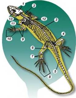
Rice. 204. The structure of the skeleton of reptiles: 1 - skull; 2 - cervical spine; 3- lumbar spine; 4 - sacral spine; 5 - tail section; 6 - femur; 7 - large and small tibia; 8 - foot; 9 - ulna and radius; 10 - brush; eleven - brachial bone; 12 - scapula; 13 - clavicle
The signs of the disease are often very similar regardless of the cause, but can be extremely subtle, especially on early stages diseases. The ability of reptiles to mask any signs of illness and the chronic nature of disease progression means that when the owner notices that the animal is not feeling well, the disease has already improved. Again, this has implications for successful treatment, therefore, if you have doubts or any of the following signs, you should seek treatment immediately.
Above: chronic pneumonia, anorexia, malnutrition, stomatitis in the Boa joint. Secondly, a detailed physical examination often gives me much more information about the patient in question than a user's description on the phone or photos and descriptions on the Internet. A detailed clinical examination is one of the main diagnostic tools that a veterinarian uses in evaluating his patient, who unfortunately cannot contact us directly as in human medicine. Observation of the breathing patterns and effort of the animals before treatment gives a good indication of the functionality and health of the respiratory tract.
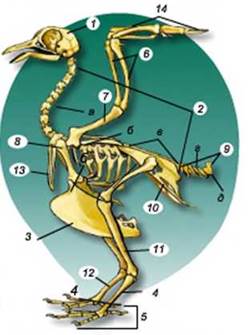
Rice. 205. Bird skeleton: 1 - skull; 2 - spine (a - cervical region; b - thoracic; c - complex buttocks; d - free tail vertebrae, e - coccygeal department); 3 - keel; 4 - handguard; 5 - foot; 6 - ulna and radius; 7 - humerus; 8 - scapula; 9 - tail vertebrae; 10 - pelvic girdle; 11 - femur; 12 - lower leg; 13 - fork (fused collarbones); 14 - brush
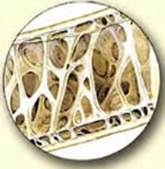
Rice. 206. Internal structure hollow bird bone
The most common way snakes move is by undulating body bends. Water snakes, sea snakes can deftly swim in the water.
The skeleton of birds (Fig. 205) is characterized by lightness, since some of the bones have cavities filled with air (Fig. 206).
It is subdivided into the skeleton of the head (skull), torso (spine and chest), limbs and their belts. characteristic feature the skull of birds is that most of its bones are fused together, and not connected with sutures, as in reptiles. A large volume of the brain box is associated with the progressive development of the brain. Attract attention and large eye sockets. The jaws of birds are elongated and, as you already know, covered with horn covers. Thanks to this, a perfect apparatus for capturing food is created, because birds have no teeth.
The spine has a number of features associated with flight. The cervical region consists of a large number of vertebrae (from 11 to 25). This gives the head considerable mobility. The vertebrae of the thoracic, lumbar, sacral sections are connected to each other motionlessly. This creates a solid foundation for the operation of the wings during flight. The complex buttocks create a reliable support for the hind limbs, since they account for the entire mass of his body. Part of the tail vertebrae remains free, and the last vertebrae fuse together, forming the coccygeal bone (Fig. 205), to which the tail feathers are attached.
You already know that in most species of birds the sternum has a flat outgrowth - the keel. Muscles are attached to it, which ensure the movement of the wings during flight. In the girdle of the forelimbs, the clavicles are fused together, forming the so-called fork (Fig. 205). It gives elasticity to the belt of the forelimbs. The wing skeleton consists of three sections: shoulder, forearm and hand. Only three fingers remain on the forelimbs of birds. The skeleton of the hind limb consists of the thigh, lower leg and foot. Most of the bones of the foot fuse together and form long bone- a trickle, which, together with the fingers, is covered with horny scales (Fig. 205). The jet gives the leg strength and increases its mobility.
The main mode of movement for most birds is flight. During takeoff, birds can use headwinds. A flight for which large and small constantly work in turn pectoral muscles, are called mahalny, or active (Fig. 207, 1). It requires a significant expenditure of muscular energy. Large birds of prey - eagles, kites, vultures - for a long time can circle in the air in search of prey, almost without moving their wings. This is a soaring, or passive, flight (Fig. 207, 2), which is carried out thanks to the ascending currents of warm air rising from the surface of the earth, unevenly heated by the sun. A soaring flight, in contrast to a flying flight, does not require a significant expenditure of energy.
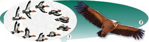
Rice. 207. Flight of birds: 1 - mahal; 2 - soaring
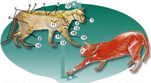
Rice. 208. A Structure of the skeleton of a mammal: 1 - skull; 2- cervical vertebrae; 3 - thoracic vertebrae; 4 - lumbar vertebrae; 5 - sacral vertebrae; 6- pelvic bones; 7 - tail vertebrae; 8 - femur; 9 - small and large tibia; 10 - bones of the foot; 11 - ribs; 12 - sternum; 13 - bones of the hand; 14 - radius; 15 - elbow bone; 18 - humerus; 17 - scapula. Would. The muscular system of a mammal (note how well differentiated it is)
Consider Figure 208. A and find on it all the components of the mammalian skeleton. Compare its structure with the skeletons of amphibians, reptiles and birds. The muscles of mammals are well developed and differentiated (Fig. 208. C). Mammals are characterized by the presence of a diaphragm, which is involved in respiratory movements: due to its reduction, the volume of the chest cavity changes. The muscles of the back, limbs and their belts are also well developed, as well as chewing muscles that move the lower jaw.
Mammals have different modes of movement (Fig. 209). Many inhabitants of the terrestrial environment, such as ungulates, leopards, cheetahs, wolves, foxes (Fig. 209, 1) are able to run. Lagomorphs (hares, rabbits) can move by jumping: therefore, they have elongated hind legs (Fig. 209, 2). Earth-dwelling mammals make their passages in the soil: moles dig with the help of their forelimbs (Fig. 209, 3), and mole rats - with the help of powerful incisors.
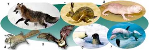
Rice. 209. Various ecological groups of mammals: 1 - ground running; 2 - jumping; 3 - land; 4, 5 - mammals with passive and active flight; 6, 7 - water forms (with the help of a teacher, identify the animals shown in the figure)
There are mammals capable of either passive or active flight. Figure 209, 4 shows a rodent - an ordinary flying squirrel. This view is from the beginning of the 20th century. lived on the territory of Ukraine. The flying squirrel spends most of its life on trees, occasionally descending from them to the ground. Between the front and hind legs, it has a skin membrane, which allows it to passively glide from tree to tree, covering a distance of up to 50 m. By changing the tension of the skin membrane, the flying squirrel is able to change the direction of flight, and the long fluffy tail serves for braking.
Active flight is inherent in rukkrylim. Remember, in these animals, from the top of the second finger of the forelimbs to the tail, a fold of skin stretches along the body, which serves as a wing (Fig. 209, 5). Like birds, chiropterans have a sternum keel and well-developed pectoral muscles that provide wing movement.
Inhabitants of water bodies, for example, cetaceans, capable of deftly swimming and diving (Fig. 209, 6). They have a streamlined body shape, the forelimbs have turned into flippers. The main organ of movement is the caudal fin.
Biological dictionary: active and passive flight, keel, tarsus.
GENERALIZATION OF KNOWLEDGE
All chordates have an internal skeleton. In vertebrates, the skeleton of the head (skull), trunk (spine, and in reptiles, birds and mammals - also the chest), limbs (includes the skeleton of the limb belts and the skeleton of the free limb) are formed.
Among vertebrates, the least differentiated muscles are in fish, and the best differentiated muscles are in terrestrial animals. This is due to the appearance of paired limbs.
Different groups of chordates have different modes of locomotion. Inhabitants of reservoirs swim in the water column, terrestrial animals crawl, run, jump, inhabitants of the soil dig passages. Birds and bats are capable of active flight.
TEST YOUR KNOWLEDGE
1. What is common and different in the structure musculoskeletal system fish and amphibians? 2. What features of the structure of the musculoskeletal system of reptiles characterize them as inhabitants of the terrestrial environment? 3. What are the features of the musculoskeletal system of birds associated with adaptation to flight? 4. What is common and different in the structure of the skeleton of birds and bats? 5. What adaptations of mammals do you know for various modes of movement?
DISCUSS IN GROUPS
How did the skeleton of vertebrates become more complex in the process of historical development?
FOR INQUIRY AND RESPONSIBLE
1. Cetaceans lack hind limbs. But sometimes cubs are born with underdeveloped hind limbs. How can this be explained? 2. Compare the features of the flight of insects and birds, as well as the structural features of their musculoskeletal system.
PRACTICAL WORK 5
Topic: Comparison of the structure of the skeletons of vertebrates
Purpose: to consider the skeletons of different representatives of vertebrates, to find similarities and differences.
Equipment, materials and objects of study: skeletons of fish, frogs, lizards, birds, mammals; rulers, drawings of skeletons of vertebrates.
Progress
1. Consider the skeleton of fish. Pay attention to the skeleton of the head (skull), the skeleton of the body (spine, limbs (paired fins and their belts).
2. Consider the skeleton of a frog. Find the head skeleton (skull), torso skeleton (spine), paired limbs and their girdles. What are the main complication of the structure of the frog skeleton compared to fish, what is the reason for this?
3. Consider the skeleton of a lizard. Find the sections of the skeleton: the skeleton of the head (skull), the skeleton of the body (spine), paired limbs and their belts. What is the shape and length of the lizard's spine? What is the structure of the spine associated with? How are the limbs located?
4. Consider the structure of the bird's skeleton. Find the departments: the skeleton of the head (skull), the skeleton of the body (spine), paired forelimbs (wings), hind limbs and their belts. What modifications did the skeleton of the fore and hind limbs of the bird undergo? What is it connected with?
5. Consider the structure of the skeleton of a mammal. Find the sections of the skeleton: the skeleton of the head (skull), the skeleton of the body (spine), paired limbs and their belts. To the skeleton of which animal is the most similar skeleton of a mammal?
6. Draw conclusions about how the structure of the skeleton depends on the lifestyle of the animal, you saw the complication in the structure of animal skeletons.