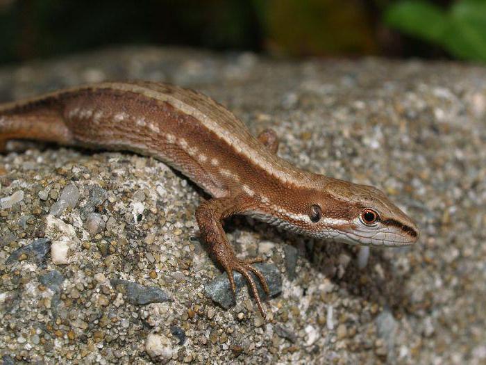
External structure reptiles consider the example of a lizard. Body lizards(as a typical representative of reptiles) is divided into sections: head, trunk, tail and two pairs of limbs (Fig. 143, BUT).
Outside, the body is covered with dense dry skin. There are no glands in the skin of a lizard. This protects the animal's body from moisture loss in an arid environment. Formed in the upper layer of the skin scales , but not bone, like in fish, but horny , softer. The growth of the body of a reptile is accompanied by molting . At the same time, the old horny cover exfoliates, bursts, and in lizards it comes off in patches. In snakes, it separates, sliding like a stocking from the whole body and is called crawling out .
The head is oval (in snakes it can be triangular) and covered with large horny shields (they even have special names). In primitive lizards, for example agam, geckos, head and torso are covered with homogeneous horny scales.
The mouth has jaws with teeth: with them the lizard grabs and holds prey. A pair of nostrils is visible above the mouth. They are transparent and let air into the mouth. Inside the nostrils are olfactory organs that lizards use to smell. A long thin tongue constantly protrudes from the mouth of lizards and snakes, serving the animal for feeling and touching surrounding objects, as well as for perceiving their smells. The lizard's eyes are covered moving eyelids .
Between the head and the body there is an interception - neck . It allows the animal to turn its head in the direction of a sound or a moving object, to grab prey and deal with it.
The body of the lizard is slightly flattened, soft. The tail is long, elastic. It can break off and then recover - regenerate . Two pairs of legs are widely spaced on the sides of the body, the toes are claws . When the lizard moves crawling - touch the body of the earth (hence the name of this class).
In connection with the terrestrial way of life and the transition to exclusively lung breathing the body of reptiles is covered with horny scales and is devoid of glands.
Skeleton. In reptiles, the skeleton, to a greater extent than in amphibians, is adapted to life on land (Fig. 143, B).
The head has one protrusion - condyle with which the back of the skull is attached to the spine. This makes the head well mobile when supported by the spine.
The spine of the lizard is divided into sections: cervical, trunk, sacral and caudal. There are 7-10 mobile vertebrae in the cervical region. The first two stand out atlas and epistrophy . Their articulation enhances the mobility of the head. To the trunk vertebrae (16-25) are attached ribs . The anterior true ribs join with the sternum and form chest . It protects the organs located in the chest cavity (esophagus, heart, lungs) from damage and participates in the breathing mechanism: it expands when inhaling and falls when exhaling.
In the skeleton of snakes, the ribs are attached to the vertebrae along the entire length of the trunk part of the spine and do not connect to the sternum (snakes do not have a chest). The pelvic girdle is attached to the sacral vertebrae (there are two of them). skeleton belts and free limbs retains the general structure of all terrestrial vertebrates. The limbs of lizards are widely spaced, but there are also legless among lizards. Snakes don't have legs either. In these cases, the reptiles move with the help of powerful muscles attached to the spine and ribs, the ends of which protrude through the skin and cling to the unevenness of the soil.
The internal structure and vital activity of reptiles
We will also consider the features of the internal structure and vital activity of reptiles using the example of a lizard.
Nutrition and digestion. Digestive systems reptiles and amphibians are similar in all major departments (Fig. 144, 145). These are the mouth, pharynx, stomach, intestines. Food gets wet in the mouth saliva which is characteristic of terrestrial animals. In the stomach under the action gastric juice in an acidic environment, protein foods are digested. The ducts of the gallbladder, liver and pancreas open into the intestine. Here digestion of food is completed, absorption of nutrients into the blood occurs.
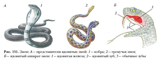
Lizards eat mainly insects and worms. The snakes prey on voles and mice. Some snakes have special sensitive pits on the front of their heads - thermolocators , able to perceive heat (infrared radiation) coming from a warm-blooded animal. Venomous snakes kill prey with venom flowing down poisonous teeth from poisonous glands located in the walls of the oral cavity.

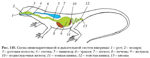
Respiratory system. In connection with the appearance of the cervical region, the lizard lengthens the airways through which air enters from the mouth into the lungs (see Fig. 145). Air is drawn in through the nostrils, enters the mouth, then larynx , then into a long tube - trachea ; The trachea divides into two even narrower tubes bronchi going to the lungs. The lungs of reptiles are more complex than those of amphibians: there are many folds inside the lung cavity, where they branch out many times. blood vessels. This increases the surface of their contact with air, increasing gas exchange.
Circulatory system. Heart three-chamber , with an incomplete septum in the ventricle. Three large vessels emerge from it: the left and right aortic arches and the pulmonary artery (Fig. 146). Two aortic arches, bypassing the heart, merge into one common vessel - the dorsal aorta.

Mixed blood flows through the body (like in amphibians), which affects fluctuating body temperature temperature dependent environment.
The pulmonary arteries carry venous blood from the heart to the lungs for oxygenation. Through the pulmonary veins, arterial blood enters the left atrium. In the ventricle, the blood is partially mixed, the most oxygenated goes to the head, mixed - to all organs of the body, saturated with carbon dioxide - to the lungs.
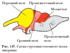
Nervous system. In reptiles, all parts of the brain become more complex and enlarge (Fig. 147) in comparison with the brain of amphibians. This is manifested in the more complex and diverse behavior of reptiles. Conditioned reflexes are formed in them faster than in fish and amphibians. The forebrain and cerebellum are especially enlarged, the medulla oblongata forms a bend characteristic of all higher vertebrates. In addition to sight and smell, reptiles have a well-developed sense of touch.
excretory system. In reptiles, the excretory system is the same as in all terrestrial vertebrates. In the excretory organs - the kidneys - the mechanism for returning water to the body is strengthened: it is absorbed by the renal tubules. Final product metabolism in reptiles is excreted not in the form of liquid urine (as in amphibians), but as uric acid in a mushy state into the cloaca, and then out. It does not require as much liquid to remove uric acid from the body as it does to remove liquid urine.
Reproductive organs. These are the testes in males and the ovaries in females (Fig. 148). Fertilization in reptiles is internal. It occurs when the cloacae of the male and female approach each other. The embryo developing in a fertilized egg, moving along the oviduct, is covered with egg and embryonic membranes. They provide the embryo with water, protect against drying, shaking, participate in respiration and excretion of metabolic products.

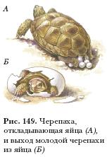
Reptiles lay their eggs on the ground or in specially prepared recesses (Fig. 149). Some reptiles guard their masonry (for example, crocodiles); others, having laid eggs, leave them (for example, turtles). Sometimes the young are carried in the mother's body. In these cases, live birth occurs, for example, in vipers and at viviparous lizard.
annual cycle of life. Reptiles are widely distributed around the globe and are found in different climatic zones. However, being cold-blooded animals with variable body temperature, they need external heating from the sun. Therefore, these animals are most numerous in the tropical and subtropical zones of the globe. In the conditions of changing seasons, when cold autumn and winter come to replace warm summer, reptiles go into shelters with the onset of adverse conditions: burrows, caves, under tree roots, under rural houses and forest huts. There animals fall into a stupor - hibernation . In the spring, when the air and the soil surface warm up well, the reptiles come to the surface and move on to active image life.
Variety of reptiles
There are over 6,000 reptiles in the class. modern species. The divisions in the class are: scaly(with suborders of lizards and snakes), crocodiles and Turtles.
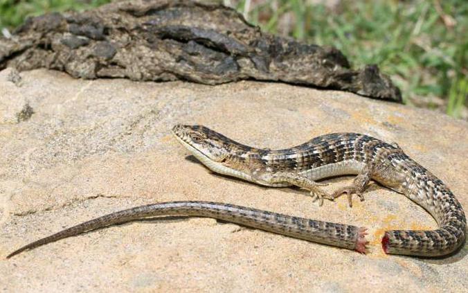
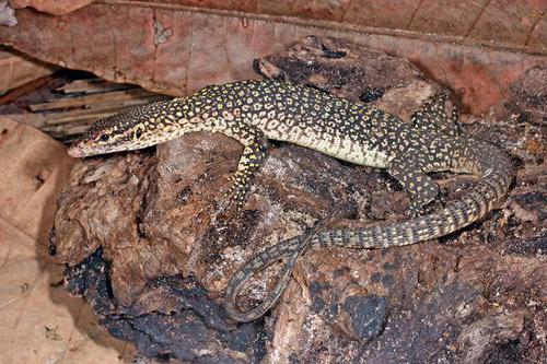
lizards they are distinguished by a flexible movable body and widely spaced legs (Fig. 150). In a temperate climate this quick and viviparous lizard, and in hot regions it is geckos, agamas, monitor lizards. known legless lizards – spindle and yellowbell. They are distinguished from snakes by non-united movable eyelids. In total, there are about 3300 species of lizards in the world. The length of the smallest lizards is about 3.5 cm, the largest - komodo dragon- more than 3 m.

snakes do not have limbs. They move thanks to the powerful muscles of the body and numerous ribs, the ends of which, appearing through the skin, cling to the unevenness of the soil. Snakes are distinguished from lizards unblinking gaze since their eyes are covered transparent horny eyelids , and the ability to crawl onto prey with a stocking thanks to moving moving jaws.
Among the snakes there are very large and strong boas, for example reticulated python, anaconda. Their length reaches 6-10 m. The boa constrictor strangles the caught victim, wrapping around the whole body. The body length of the smallest snakes is no more than 8 cm.
A lot of poisonous snakes: cobra, gyurza, viper, rattlesnake, efa. They kill the victim with the poison of poisonous teeth (Fig. 151). Poisonous snakes are also dangerous to humans. Their bites cause serious illness and even death. In medicine, remedies are known to avoid the serious consequences of snake bites.
First aid measures in cases of a venomous snake bite, the following: splinting, calm position of the damaged organ, plentiful warm drinking. The most effective is the introduction of an anti-snake serum preparation.
The main way to avoid being bitten is to be very careful when moving and stopping in areas where many poisonous snakes live. Snakes do not like to use poison for defense, they need it as a means of obtaining food. Therefore, with the approaching noise, they tend to hide.
People have learned to use snake venom for medical purposes and use it to treat many diseases.
To non-venomous snakes relate snakes, snakes, boas. They grab prey with their teeth and then swallow it.
About 2,700 modern species of snakes are known, about a third of them are poisonous.
crocodiles- large and strong reptiles living in tropical countries (Fig. 152). Their body length reaches 6 m. They live along the banks of rivers and lakes, hunt for prey in the water. A hidden crocodile grabs a large animal (for example, an antelope) that has come to a watering place. Crocodiles are good swimmers, using a long, laterally compressed tail and webbed feet. The body of the crocodile is completely immersed in water, and only the nostrils and eyes located on the elevations of the skull remain above the surface.
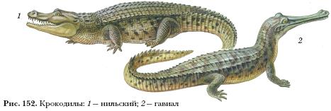
Among crocodiles there are alligators, real crocodiles, gharials, caimans. In total, there are 21 species in the world.
Turtles- the most ancient group among reptiles. Their appearance is very specific: the body is hidden under a powerful hard bone shell (Fig. 153).
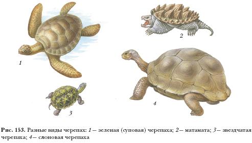
Turtles do not molt, so the dark and light annual stripes on the horny shell plates can determine their age. Although turtles move slowly on land, they are difficult for predators to reach, as they pull their heads and legs under their shells when threatened.
Turtles include terrestrial, freshwater and marine species. Their movements in the water are very fast and maneuverable. The largest turtles are marine, for example green (soup) turtle up to 150 cm long and weighing up to 400 kg. Of the land turtles, the largest - Galapagos elephant tortoise, with a shell 150 cm long and weighing up to 400 kg. In total, more than 200 modern species of turtles are known in the world.
Many species of crocodiles and turtles have become very rare, they need protection and are listed in the Red Books.
The value of reptiles. ancient reptiles
The value of reptiles. Most lizards and snakes, eating insects that harm agriculture, rodents and terrestrial mollusks, bring benefits to humans. In some countries in South America, South Asia and Africa, non-venomous snakes are kept instead of cats. In nature, reptiles are woven into a common system food links : some turtles eat plants, others - animals (insects, amphibians, reptiles, small animals), and they, in turn, are eaten by other predators - birds of prey and animals.
Sometimes land turtles cause damage to melons, water snakes - to fish farms. Reptiles can spread pathogens to humans and domestic animals.
Poisonous snakes are dangerous with their bites. At the same time, the study of snake venom led to the creation of valuable medicinal preparations based on them, which people use for diseases of the respiratory organs, heart, and joints.
Large snakes and crocodiles are harvested to make beautiful and durable leather. Sea turtles are hunted because of the delicious meat. Because of this, the number of many species has sharply decreased, and some are on the verge of extinction. Reserves have been created for them. Listed in the IUCN Red List Galapagos elephant tortoise, green tortoise, Komodo dragon, Cuban crocodile, tuatara.
Eating plants, insects, amphibians, small animals, reptiles are consumers of ready-made organic substances. Among them there are herbivorous and insectivorous, but the majority are predatory (carnivorous).
Ancient reptiles. Modern reptiles evolved from ancient amphibians - stegocephalians who lived in the middle of the Paleozoic era. The most ancient of reptiles are considered cotylosaurs who lived 230-250 million years ago. Some features of their organization are preserved in the form of turtles.
The heyday of reptiles was the Mesozoic era (250-65 million years ago). In those ancient times, they lived on land and in water, flew in the air (Fig. 154).

flying pterodactyls, rhamphorhynchus, pteranodon looked like giant bats. Their wingspan reached 10-12 m. Lizards similar to dolphins and seals lived in the water. These were ichthyosaurs, plesiosaurs. These groups of ancient reptiles died out without leaving any descendants.
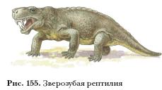
Among the ancient lizards there were two more groups that played important role in the appearance of birds and mammals: dinosaurs and animal-like reptiles(Fig. 155).
Dinosaurs were a very diverse group: peaceful (herbivorous) and ferocious predators. Some moved on four legs, others only on two hind legs, in an upright position. Very large dinosaurs are also known - more than 30 m long, and small ones - the size of a small lizard. Considered the largest diplodocus(length 27 m and weight about 10 tons), Apatosaurus, Brachiosaurus, Seismosaurus. They lived near water bodies and stood in the water for a long time, eating aquatic and near-water vegetation. Some dinosaurs had combs on their backs that they used to capture solar energy. Scientists suggest that birds originated from one of the groups of dinosaurs.
 Animal reptiles
got their name for their resemblance to animals (see also § 51) . Unlike other lizards, their legs were located under the body, lifting it off the ground. Fangs stood out among their teeth, fleshy lips
, a skin probably had glands.
Animal reptiles
got their name for their resemblance to animals (see also § 51) . Unlike other lizards, their legs were located under the body, lifting it off the ground. Fangs stood out among their teeth, fleshy lips
, a skin probably had glands.
However, throughout the Mesozoic era, the fate of dinosaurs and animal-like reptiles was different. Dinosaurs were favored by the warm, mild climate of this era and dominated everywhere. The bestials were few in number and invisible. At the end of the Mesozoic era, the ratio of the number of species began to change in favor of animal-like ones.
The extinction of dinosaurs occurred when the climate of the planet changed, since at the end of the Mesozoic, a long warm period was replaced by low temperatures. At this time, vegetation began to change, and with the beginning of the Cenozoic era, angiosperms began to spread on Earth.
Many scientifically proven (mountain building and climate change) and alleged reasons for the extinction of dinosaurs are put forward. It is possible that a large asteroid passed near the Earth, which influenced climate change and the natural environment surrounding dinosaurs.
Did the ancient lizards disappear from the face of the planet without a trace, leaving only monuments in the form of skeletons and prints? In the modern fauna of reptiles there are tuatara, which is called living fossil . There is a lot of archaic in the appearance of this animal: the remains of a shell on the body, the primitive structure of the spine, an additional eye in the parietal part of the head. This reptile lives on small islands near New Zealand and is strictly protected as a living monument of nature. Tortoises are close to their Mesozoic ancestors.
According to some features of the organization, crocodiles are close to dinosaurs.
Lizards and snakes also have certain similarities with dinosaurs. But in the history of the fauna of the Earth's vertebrates, they appeared only in the Cenozoic era, when their related groups lost their former greatness.
Axial skeleton of reptiles. In reptiles, compared to amphibians, axial skeleton or spine, is divided into departments more distinctly. cervical consists of several vertebrae. The two anterior cervical vertebrae have a special arrangement. One is called an atlas. This vertebra differs from the rest in that it does not have a vertebral body, and it has the shape of a ring divided into 2 parts. The second cervical vertebra is called the epistrophy. This vertebra has a large process in front, which is shaped like a tooth. Due to this structure of the cervical vertebrae, reptiles have a very mobile head. Other cervical vertebrae have a normal structure.
The lumbar and thoracic regions of reptiles are practically the same, therefore they are usually considered as one whole department. The thoracic region of the reptile skeleton is considered to be only that part of the spine in which the ribs extend from the vertebrae and are attached to the sternum. In the lumbar region, the vertebrae do not have ribs that reach the sternum. The vertebrae of reptiles are concave in front and convex in the back. They are called targeting. Arches rise above the vertebra, which end with the axillary process. The upper arcs form a special channel in which the spinal cord. Articular processes extend from the base of the upper arches, also anterior and posterior. These processes of the upper arches are connected to the processes that were formed from neighboring vertebrae. These connections contribute to greater spine strength when the reptile flexes. There are small indentations on the vertebral body on the sides, ribs join them.
The sacral section of reptiles consists of 2 vertebrae. Their main characteristics are very well developed transverse processes, to which the pelvic bones are attached. The tail section of reptiles consists of numerous vertebrae, which gradually decrease in size.
It is this structure of the spine that is characteristic of many reptiles. But there are some species in which the spine is somewhat modified. For example, in snakes, the spine is divided only into the tail and trunk sections, since they do not have paired limbs, and they have a different type of movement - as you know, snakes crawl on their belly, bending their bodies. All trunk vertebrae of snakes have movable ribs. The lower ends of the ribs are free, and the sternum is absent.
Scull. If we compare the skull of amphibians and reptiles, then the latter have a more ossified skull. But there is also a small amount of cartilage, which is found only in the auditory region and the olfactory capsule. Visceral and axial departments skulls are laid separately even when the snake is in the embryonic stage. But, as the snake grows older, they become one. The skull consists of cartilaginous and numerous skin bones. Scientists very often use the skull of the largest lizard, the monitor lizard, to study this part of the body of reptiles. So, now you know that the structure of the skeleton of reptiles and amphibians are different from each other.
What parts of the reptile brain are the most developed?
The cerebral hemispheres are larger than those of amphibians; they have a very thin, gyri-free cortex of gray medulla. The cerebellum is highly developed. The medulla oblongata forms a pronounced bend in the vertical plane, which is characteristic of all higher vertebrates. 12 pairs of cranial nerves leave the brain.
The structure of the cervical spine of reptiles
What are the structural features of the cervical spine of reptiles?
In the cervical region, the lizard has 8 vertebrae. A feature of this department of the axial skeleton is the formation of a joint between spinal column and skull and, therefore, providing greater mobility of the head. The first cervical vertebra is a bone ring divided by a ligament into two holes: the spinal cord passes through one, the odontoid process of the second cervical vertebra enters the other, around which the first vertebra rotates.
Sections of the spine of reptiles
What are the parts of the reptile spine?
The reptile spine consists of the cervical, thoracic, sacral, and caudal sections.
horn cover of reptiles
What is the meaning stratum corneum reptiles?
The horny cover protects animals from moisture loss and desiccation, protects from mechanical damage.
The external structure of reptiles
What are the features of the appearance of reptiles?
Reptiles are the first true terrestrial vertebrates.
General features of appearance for reptiles include the following:
- the body is divided into head, neck, torso (chest and belly) and tail;
- two pairs of limbs are placed on the sides of the body so that the hips and shoulders are parallel to the ground, as a result of which the body is very slightly raised above the ground, and the animal moves, characteristically wriggling its body and almost dragging its belly along the ground, i.e., crawling;
- at the ends of the fingers there are horny claws;
- the skin is dry, devoid of glands, covered with horny shields (protection from drying out); for many representatives, for example snakes, molting is characteristic - periodic shedding of the horny cover.
reptile evolution
Who are the ancestors of reptiles?
Reptiles descended from ancient amphibians - stegocephals, as evidenced by numerous similarities in their structure, in particular, the presence of a parietal, third eye in reptiles, which is also typical of ancient lobe-finned fish and amphibians.
The value of amphibians
What is the role of amphibians in nature and in human life?
Amphibians occupy an important place in food chains. Eating mainly animal food, they destroy a huge number of insects - pests of agriculture and carriers of pathogens of humans and animals, such as mosquitoes, mosquitoes, etc. Many toads in the middle lane, settling in fields and gardens, exterminate slugs and other pests. But amphibians also cause harm by eating fish fry of valuable species.
Amphibians themselves serve as food for many birds (ducks, cranes) and mammals (black polecat, raccoon dog).
In some countries, the population eats the meat of large frogs and salamanders.
Amphibians are used for experiments as laboratory animals.
Cloaca
What is a cloaca?
Cloaca (from lat. cloaca - a pipe for draining sewage) - an expanded end part of the hindgut that opens outwards. The ureters and genital ducts open into the cloaca.
Reproduction and development of amphibians
How is reproduction and development of amphibians carried out?
Sexual maturity in many frogs occurs at three years. In spring, males begin to croak; some species have resonators for this. The females lay their eggs in the water, and the males fertilize them. The fertilized eggs then float to the surface, where the water is warmer. The eggs are dark above (to trap heat), and light below. The egg is completely, but unevenly crushed. After 8–10 days (in frogs) after fertilization, the embryo breaks through the egg membranes, and the larva (tadpole) comes out. Initially, the tadpole does not have paired limbs, and the function of the organ of movement is performed by the tail, bordered by a well-developed membrane. It has many signs of fish: one circle of blood circulation, a two-chambered heart, a lateral line, external gills, which then change to internal ones. First, the tadpole lives off the supply of eggs, and then it develops a mouth with horny jaws, and it feeds on algae and sessile protozoa, scraping them from plants and from the bottom. The hind legs appear first, then the front legs, and the lungs develop. As they develop, the internal gills disappear, the circulatory system changes, the intestines shorten, and other systems change. It shortens, and then the tail disappears completely. From the moment of fertilization to the appearance of a frog, 2–3 months pass, depending on the ambient temperature.
The structure of the circulatory system of amphibians
What is the structure circulatory system amphibians?
The circulatory system has two circles of blood circulation. The three-chambered heart of amphibians consists of one ventricle and two atria. Both atria and then the ventricle contract alternately. The right atrium receives venous blood from the organs and tissues of the body (from great circle blood circulation), to the left - arterial blood from the lung (from the pulmonary circulation). In the ventricle, the blood mixes, but only partially, due to the presence of special distribution mechanisms (spiral valve, outgrowths and pockets) that prevent mixing of blood portions coming from different atria into the ventricle. The brain receives oxygen-rich arterial blood, which flows through carotid arteries coming out of the heart. The trunk and limbs are supplied with mixed blood coming through the aortic arches. The oxygen-depleted blood enters the pulmonary arteries (the pulmonary circulation), is enriched in the lungs with oxygen, and enters the left atrium in the form of arterial blood.
The low rate of blood flow and mixing of blood in the ventricle is evidence of a low metabolic rate. Amphibians, like fish, are cold-blooded animals, that is, their temperature depends on the ambient temperature.
Lizards, being a suborder of the class of reptiles, are its most numerous group. These reptiles number more than 3,500 species and live on all continents except Antarctica. In this article, we will look at internal structure, skeleton, physiological features of the lizard, species and names of their families.
Lizards are amazing creatures, which are distinguished from the rest of the fauna by several interesting facts. The first fact is the size of representatives of different populations of lizards. So, for example, the smallest lizard Brookesia Micra is only 28 mm long, while the largest representative of this group of reptiles, the Indonesian monitor lizard, also known as the Komodo dragon, has a body length exceeding 3 meters, with a weight of about one and a half centners.
The second fact that makes these reptiles popular not only among biologists, but also ordinary people, is why and how the lizard sheds its tail. This ability is called autotomy and is a method of self-preservation. When a lizard runs away from a predator, he can grab her by the tail, which actually poses a threat to the life of the reptile. In order to save their lives, some species of medium-sized lizards are able to shed their tail, which grows again after a while. To avoid large blood loss during autotomy, the tail of the lizard is equipped with a special group of muscles that reduce blood vessels.
In addition to all that is listed above, lizards in nature have the quality of skillful disguise, adapting to the color scheme of the environment. And some of them, especially the chameleon, can take on the color of an adjacent object in a matter of moments. How does this happen? The fact is that the skin cells of a chameleon, consisting of several almost transparent layers, have special processes and pigment, which, under the influence of nerve impulses may shrink or expand. At the moment of contraction of the process, the pigment is collected in the center of the cell and becomes barely noticeable, and when the process is unclenched, the pigment spreads throughout the cell, staining the skin in a certain color.
Skeleton and internal structure of a lizard
The body of a lizard consists of such parts as the head, neck, torso, tail and limbs. The body is covered with scales on the outside, consisting of smaller and softer horn formations compared to fish scales, there are no sweat glands on the skin. A characteristic feature is also a long muscular organ - the tongue, which is involved in feeling objects. The eyes of a lizard, unlike other reptiles, are equipped with a movable eyelid. Musculature has a greater degree of development than that of reptiles.
The lizard skeleton also has some features. It consists of the cervical, shoulder, lumbar and pelvic regions, which are connected by the spine. The skeleton of the lizard is built in such a way that, when fused, the ribs (the first five) form a closed sternum from below, which is characteristic feature this group of reptiles compared with other reptiles. The chest performs protective function reducing the risk of mechanical damage internal organs, it can also increase in volume during breathing. The limbs of the lizard, like those of other terrestrial vertebrates, are five-fingered, but unlike amphibians, they are located in a more vertical position, which provides some elevation of the body above the ground and, as a result, faster movement. Significant assistance in movement is also provided by the long claws with which the paws of the reptile are equipped. In some species, they are especially tenacious and help their master deftly climb trees and rocky terrain.
The lizard skeleton differs from other groups of terrestrial representatives of the fauna by the presence of only 2 vertebrae in the sacral spine. Also, a distinctive feature is the unique structure of the caudal vertebrae, namely in the non-ossifying layer between them, due to which the lizard's tail is painlessly torn off.
What are the similarities between a lizard and a newt?
Some people confuse lizards with newts - representatives of the infraorder. What are the similarities between a lizard and a newt? Representatives of these two superclasses are similar to each other only externally, the internal structure of newts corresponds to the anatomy of amphibians. Nevertheless, from the point of view of physiology, both lizards and newts visually look the same: a snake-like head, movable eyelids on the eyes, a long body with five-fingered limbs on the sides and sometimes with a crest on the back, a tail capable of regeneration.
Lizard food
The lizard belongs to cold-blooded animals, that is, its body temperature changes depending on the ambient temperature, so these reptiles are most active during the day, when the air warms up the most. Most of them are carnivorous lizards, the species and names of which include more than one thousand individuals. The prey of predatory lizards directly depends on the size of the reptile itself. So, small and medium-sized individuals feed on all sorts of things such as insects, spiders, worms, mollusks. The victims of large lizards are medium-sized vertebrates (frogs, snakes, small birds or lizards). The exception is the Komodo monitor lizard, which, due to its large size, can afford to hunt larger game (deer, pigs, and even medium-sized buffaloes).
Another part of the lizards is herbivorous, eating leaves, shoots and other vegetation. However, there are also omnivorous species, such as Madagascar geckos, which eat plant foods (fruits, nectar) along with insects.
Lizard classification
The variety of lizards is quite impressive and includes 6 superfamilies, subdivided into 37 families in total:
- Iguanas.
- Geckos.
- Skinks.
- Fusiform.
- Monitor lizards.
- Worm-like.
Each of these infraorders has initializing features determined by the conditions of the habitat and the intended role in the trophic chain.
iguanas
Iguanas are an infraorder with many varieties of life forms, in which not only the external, but often the internal structure of the lizard differs. Iguana likes include such well-known families of lizards as the iguana, agamo and chameleon family. Iguanas prefer a warm and humid climate, so their habitat is the southern part of North America, South America, as well as some tropical islands (Madagascar, Cuba, Hawaii, etc.).

Representatives of the infraorder iguanas can be recognized by the characteristic strongly elongated lower jaw due to pleurodont teeth. Also distinctive feature iguanas is the presence of a spiny crest on the back and tail, the size of which is usually larger in males. The paw of the iguana lizard is equipped with 5 fingers, which are crowned with claws (in arboreal species, the claws are much longer than those of terrestrial representatives). In addition, iguanas have helmet-like growths on their heads and throat pouches that serve as a threat signaling tool and also play an important role in mating.
The body shape of iguanas is predominantly of two types:
- A high body with compressed sides, which smoothly passes into a thickened tail. This body shape is mainly found in tree-dwelling individuals, such as the genus Polychrus in the South American range.
- A flattened disc-shaped body - found in representatives of iguanas living on earth.
geckos
The gecko-like infraorder includes the Cepkopale, Scale-footed and Eublepharidae families. The main and common feature of all representatives of this infraorder is a special chromosome set and a special muscle near the ear. Most geckos do not have a zygomatic arch, and their tongue is thick and not forked.
- The family of gecko (chain-toed) lizards has been living on Earth for more than 50 million years. The lizard's skeleton and physiological features are adapted for living all over the world. They have the most extensive habitat both in hot climatic zones and in temperate latitudes. The number of species of the family is more than a thousand.
- The Scalefoot family is one of the outwardly very reminiscent of snakes. You can distinguish them from snakes by the characteristic clicking sound that they are able to make to communicate with each other. The body, like that of snakes, is long, smoothly turning into a tail, which is adapted for autotomy. The head of the lizard is covered with symmetrical shields. The population of Cheshuenogs includes 7 genera and 41 species. Habitat - Australia, Guinea and nearby land areas.
- The Eublepharidae family are small lizards about 25 cm long with a variegated color, leading a nocturnal lifestyle. Carnivores, feed on insects. They live on the American, Asian and African continents.
Skinks
Representatives of skink-like lizards are common on all continents with a temperate, tropical and subtropical climate. These are mainly land dwellers, although there are also semi-aquatic individuals, those who spend a greater period of their lives on trees. This infraorder includes the following families:
Spindle Lizards
The infraorder of fusiform lizards is characterized by small scales with bone plates not fused together from below. Among the spindle-shaped lizards, there are both legless species and lizards with the usual body structure with five-fingered limbs. The infraorder includes three families:
- The Xenosaur family differs from other families in its developed limbs and heterogeneous scales. Highlights the presence of movable eyelids and auditory openings. The family includes only two genera with habitats in Central America and China.
- The spindle family has strong jaws equipped with blunt teeth. Basically, these are carnivorous lizards that reproduce by live birth. The family includes about 10 genera and 80 species, living mainly on the American continent. The size of adults ranges from 50-60 cm.
- The Legless family has only two species with a habitat in Mexico and California. They are distinguished by the absence of limbs, auditory openings and bone plates.

monitor lizards
The infraorder Varaniformes includes one genus - Monitor lizards - and about 70 species. Monitor lizards live in Africa, with the exception of Madagascar, Australia and New Guinea. The largest species of monitor lizards, the Komodo monitor lizard, is a real champion among all types of lizards in size, its length reaches 3 meters and its weight is more than 120 kg. His supper could very well be a whole pig. The smallest species of monitor lizards (Short-tailed Monitor) does not exceed 28 cm in length.
Description of the monitor lizard: an elongated body, an elongated neck, limbs in a semi-straightened position, a forked tongue. Monitor lizards are the only genus of lizards in which the skull is completely ossified, there are open ear holes on the sides. The eyes are well developed, equipped with a round pupil and a movable eyelid. The scales on the back consist of small oval or round plates, on the belly the plates take on a rectangular shape, on the head they are polygonal. A powerful body ends with a no less powerful tail, with which monitor lizards are able to defend themselves, inflicting strong blows on the enemy. In aquatic lizards, the tail is used to balance when swimming; in arboreal species, it is quite flexible and tenacious, helping to climb branches. Monitor lizards differ from most other lizards in the structure of the heart (four-chambered), similar to mammals, while the heart of a lizard from other infraorders has three chambers.
In terms of lifestyle, terrestrial species predominate among monitor lizards, but there are also those that spend a lot of time in the water and on trees. The body of the lizard is adapted to living in various biotopes, they can be found in the desert, and in humid forests, and on the sea coast. Most of them are predators, active in daytime days, only two species of monitor lizards are herbivorous. Various mollusks, insects, fish, snakes (even poisonous!), birds, reptile eggs, and other types of lizards become prey for carnivorous lizards, and large monitor lizards often become cannibals, eating their young and immature relatives. The entire genus of monitor lizards belongs to the oviparous lizards.
Monitor lizards are important not only as a link in the food chain for their habitat, but also for anthropological activities. Thus, the skin of these lizards is used in the textile industry as a material for the manufacture of various haberdashery and even shoes. In some states, the local population eats the meat of these animals for food. In medicine, monitor lizard blood is used to make antiseptics. And, of course, these lizards often become inhabitants of terrariums.
worm-like lizards
The infraorder of worm-like lizards consists of one family, the representatives of which are small, legless individuals, outwardly similar to worms. They live on the ground and lead a burrowing lifestyle. Distributed in the forest zone in Indonesia, the Philippines, India, China, New Guinea.
Axial skeleton. The differentiation of the axial skeleton, or spine, into sections is much more pronounced in reptiles than in amphibians. The cervical region is always composed of several vertebrae, of which the two anterior ones have a special arrangement. The first cervical vertebra is called the atlas or atlas. It is devoid of a vertebral body and has the shape of a ring divided into two parts. On the lower anterior surface of this vertebra there is an articular cavity, movably connected to the condyle of the skull. The second cervical vertebra - the epistrophy, has a large odontoid process in front, which is the body of the first cervical vertebra, fused with the epistrophy. The odontoid process freely enters the lower opening of the atlas. This structure of the first cervical vertebrae provides greater mobility of the head. Rest cervical vertebrae have a conventional device; many of them bear short cervical ribs.
.
A - atlas; B - epistrophy; B - thoracic vertebra;
D - longitudinal section of the thoracic vertebra:
1 - odontoid process of the epistrophy, 2 - vertebral body, 3 - upper arch,
4 - spinous process, 5 - canal for the spinal cord, 6 - anterior articular process,
7 - posterior articular process
The thoracic and lumbar regions are not clearly distinguished and are usually considered as a single department. Actually, the thoracic region is considered to be that part of the spine in which the ribs extending from the vertebrae are attached to the sternum with their lower end. Vertebrae lumbar bear ribs that do not reach the sternum. The vertebral bodies are concave anteriorly and convex posteriorly; such vertebrae are called procoelous. Above the vertebral body, the upper arches rise, ending with the spinous process. The spinal cord is located in the canal formed by the superior arches.
The anterior and posterior articular processes, respectively, depart from the anterior and posterior sections of the base of the superior arches. These paired processes connect with the articular processes of neighboring vertebrae and contribute to greater strength of the spine during bending. On the sides of the vertebral body (near the base of the upper arches) there are small depressions to which the ribs are attached.
The sacral section consists of two vertebrae, which are characterized by powerfully developed transverse processes; they are joined by the bones of the pelvis. The tail section is represented by numerous vertebrae, gradually decreasing in size.
This structure of the spine is typical of the class of reptiles, but in some groups it undergoes secondary changes. In particular, in snakes, due to the reduction of paired limbs and the emergence of a different type of movement - crawling on the belly by bending the body - the spine is clearly divided only into the trunk and tail sections. All trunk vertebrae have movable ribs, the lower ends of which are free (the sternum is absent in snakes) and rest against the ventral horny scutes.

.
A - carapace; B - plastron:
1 - trunk section of the spinal column, 2 - costal plates,
3 - marginal plates, 4 - coracoid, 5 - scapula, 6 - ilium,
7 - pubic bone, 8 - ischium
In turtles, the axial skeleton takes part in the formation of the bone base of their shell. The upper shield of the shell - the carapace - is composed of several rows of bone plates. The middle (unpaired) row of these plates is formed by the fusion of the expanded and flattened spinous and transverse processes of the trunk vertebrae with the skin bones; on the sides of the middle row are paired rows of bone plates fused with expanded ribs. The edge of the carapace is formed by bone plates of integumentary origin. Thus, the thoracic spine of turtles is motionless and firmly fused with the dorsal shield of the shell. The cervical and caudal sections of the spine are mobile. At the same time, the anterior cervical vertebrae are opisthocoelous (the body of the vertebra is convex in front, concave in the back), the posterior ones are longitudinal, and between these two groups there is one vertebra, the body of which has a convex surface both in front and behind.
Scull. Compared with amphibians, the skull of reptiles is characterized by a much more complete ossification. Some cartilage is preserved only in the olfactory capsule and in the auditory region. The axial and visceral regions of the skull are embryonicly laid down separately, but in adult animals they are unified education. The structure of the skull includes both cartilaginous (replacing, or primary) and numerous skin (integumentary, or secondary) bones. It is convenient to use the skull of a large lizard - monitor lizard as the main object for study.
.
A - on the side; B - from below; B - from above; G - behind:
1 - main occipital bone, 2 - lateral occipital bone,
3 - superior occipital bone, 4 - large occipital foramen,
5 - occipital condyle, 6 - anterior bone,
7 - main sphenoid bone, 8 - vomer, 9 - parietal bone,
10 - frontal bone, 11 - nasal bone, 12 - prefrontal bone,
13 - preorbital bone, 14 - lacrimal bone, 15 - superior temporal fossa,
16 - postfrontal bone, 17 - squamous bone, 18 - premaxilla,
19 - maxillary bone, 20 - zygomatic bone, 21 - rupture of the lower temporal arch due to reduction of the quadratojugal bone, 22 - square bone,
23 - pterygoid bone, 24 - palatine bone, 25 - upper pterygoid bone,
26 - transverse bone, 27 - surangular bone, 28 - dentary, 29 - angular bone,
30 - articular bone, 31 - coronoid bone
Axial skull
. In the occipital region of the skull there are all four occipital bones: the main occipital, two lateral occipital and superior occipital. These primary bones surround the foramen magnum. The lower and lateral occipital bones together form the only (unlike amphibians) occipital condyle, movably articulating with the first cervical vertebra - atlas. The articulation of the head with the neck with the help of only one condyle, in combination with the already considered structural features of the first two cervical vertebrae, gives the reptile head considerable mobility.
In the auditory region of the cartilaginous bones, only the paired anterior ear bone retains its independence, while the upper ear bones fuse with the superior occipital bone, and the posterior ear bones with the lateral occipital bones.
The interorbital septum in reptiles is thin, membranous, and only in crocodiles and lizards does it have separate small ossifications, apparently corresponding to the oculo-sphenoid bones. The olfactory capsule has no ossifications.
At the base of the skull, in front of the main occipital bone, there is a rather large integumentary main sphenoid bone. Its anterior narrow process is homologous to the parasphenoid (parasphenoideum), which is markedly reduced in reptiles. In the anterior part of the bottom of the skull, under the olfactory region, there is a paired vomer, which also has an integumentary origin.
The roof of the skull is represented by numerous integumentary bones, some of which descend downward and cover the skull from the sides. These include the parietal, frontal, and nasal bones. In front of the frontal bones, paired prefrontal and preorbital bones are usually located, and under them in the anterior wall of the orbit are paired lacrimal bones perforated by a narrow canal.

A - from above; B - bottom:
1 - premaxilla, 2 - maxillary bone, 3 - zygomatic bone,
4 - square-zygomatic bone, 5 - square bone, 6 - external nostril,
7 - orbit, 8 - lateral temporal fossa, 9 - superior temporal fossa, 10 - squamous bone,
11 - postfrontal (postorbital) bone, 12 - parietal bone, 13 - frontal bone,
14 - prefrontal bone, 15 - nasal bone, 16 - lacrimal bone, 17 - palatine bone,
18 - pterygoid bone, 19 - transverse bone,
20 - choanae (internal openings of the nostrils), 21 - occipital condyle
Of the other integumentary bones of the axial skull, the bones that take part in the formation of the so-called temporal arches are of particular interest. In a crocodile in the roof of the skull outward from the parietal bone on each side there is a hole - the upper temporal fossa. Along the outer edge, the superior temporal fossa is bounded by the postfrontal or postorbital squamous bones. These two bones together make up the superior temporal arch. On the side of the skull behind the orbit are the lateral temporal fossae, bounded from the outside by the inferior temporal arches. Each lower temporal arch is composed of two bones: the zygomatic and quadratojugal. The lower temporal arch is connected to the upper jaw: the zygomatic bone grows to the maxillary, and the quadrato-zygomatic to the square. This type of skull, like that of a crocodile - with two temporal pits and two temporal arches, is called diapsid (double-arc).
In the monitor lizard, the superior temporal fossa is limited by the complete superior temporal arch. In the composition of the inferior temporal arch, the quadratojugal bone was reduced and only the zygomatic bone was preserved; the lateral temporal pits are consequently not closed from the outside and remain open. Therefore, the skull of the monitor lizard can be considered as a skull of the diapsid type, but with a reduced lower arch. In some other lizards, the superior temporal arch is also partially reduced, while in snakes, both temporal arches are reduced (the postfrontal and squamosal bones do not connect to each other; both temporal fossae remain open on the outside). Thus, snakes and lizards (squamous order, Squamata), according to the structure of the skull, belong to the group of diapsid (two-arc) reptiles, but are characterized by varying degrees of reduction of the temporal arches.
:
1 - false temporal fossa, 2 - premaxilla, 3 - maxillary bone,
4 - zygomatic bone, 5 - square-zygomatic bone, 6 - square bone,
7 - squamous bone, 8 - postfrontal bone, 9 - parietal bone,
10 - frontal bone, 11 - prefrontal bone, 12 - upper occipital bone
In a turtle, both temporal fossae are absent, and the lateral wall of the roof of the skull, which delimits a large cavity from the outside - the so-called false temporal fossa, formed as a recess in the occipital part of the skull, is composed of densely fused bones: posterior frontal, squamous, zygomatic and square-zygomatic. This type of skull, devoid of true temporal pits and the temporal arches limiting them, is called anapsid (archless).
Visceral skull . In the monitor lizard, the palatine-square cartilage ossifies, forming a square bone in the posterior section, to the lower end of which it is attached lower jaw; the upper end of the quadrate is movably articulated with the axial skull. In front of the square bone is the pterygoid bone, and in front of it is the palatine bone, which is connected to the maxillary bones and the vomer. All these bones are paired; of these, only the quadrate bones are of cartilaginous (primary) origin.
The superior pterygoid extends upward from the pterygoid bone. This paired bone, connecting the pterygoid and parietal bones, is homologous to the vertical ("ascending") process of the palatine-square cartilage and is characteristic of living reptiles for lizards and tuatara. In addition to the upper pterygoid bones, the transverse bones depart from the pterygoid bones, which in their front part are attached to the maxillary bones. Secondary upper jaw represented by the premaxillary and maxillary bones. The lower jaw consists of a primary articular bone and integumentary bones: dentary, angular, supraangular, coronoid, and, sometimes, several other small bones.
The premaxillary, maxillary, and dentary bones of reptiles (except turtles) have simple conical, sometimes slightly recurved teeth, which adhere to the edge of the corresponding bone.
The hyoid arch, like that of amphibians, has completely lost its suspension function. The upper element of the hyoid arch (hyomandibular) is part of the middle ear in the form of a rod-shaped auditory ossicle - the stirrup (stapesseucolumella), and the rest of it, together with the remnants of the anterior branchial arches, forms the hyoid apparatus.
The described structure of the visceral skull is generally typical of all reptiles. But in some groups there are deviations from this scheme, connected mainly with the specifics of the biology of these groups.
:
1 - premaxilla, 2 - maxillary bone. 3 - palatine bone,
4 - pterygoid bone, 5 - transverse bone, 6 - square bone, 7 - squamous bone,
8 - postfrontal bone, 9 - poisonous tooth, 10 - frontal bone, 11 - nasal bone,
12 - dentary. 13 - angular bone, 14 - articular bone
In snakes, not only the square, but also the squamous bones connected to them, as well as the pterygoid and palatine bones, are very mobile. The last two carry sharp teeth. The transverse bones in snakes serve as levers that transmit the movements of the pterygoid bones to the maxillary bones, which in turn are very mobile. This whole system of movably articulated bones not only contributes to an extremely wide opening of the mouth, but also provides independent movements of the right and left halves of the jaw apparatus when pushing the prey into the pharynx with alternate interception. This allows snakes to swallow relatively very large (exceeding the thickness of the snake's body) prey. In poisonous snakes, on the maxillary bones, there are movably attached sharp, backward-curved poisonous teeth, which have an internal channel or groove on the front surface, through which, when bitten, poison flows into the wound from poisonous glands located at the base of the tooth.
The skull of crocodiles is characterized by the fact that the teeth do not adhere to the edge of the dentary, premaxillary and maxillary bones, as in other reptiles, but sit in special depressions (holes, or alveoli) of these bones - thecodont teeth. Another feature of the visceral skull of crocodiles is the secondary hard palate that separates the oral cavity from the nasopharyngeal passage. The palatine processes of the premaxillary and maxillary bones, as well as the palatine and pterygoid bones, take part in the formation of the secondary hard palate. Due to the formation of the hard palate, the secondary choanae are carried back and located in pterygoid bones, above the larynx. The formation of a secondary hard palate is associated with the nature of the lifestyle of crocodiles: direct contact of the larynx with the choanae opens up the possibility of uninterrupted breathing when eating and when the crocodile is resting in shallow water, exposing its nostrils from the water, while oral cavity filled with water.
Paired limbs and their belts. The shoulder girdle of reptiles consists of typical bones: a scapula located more dorsally and a coracoid facing ventrally. Both of these bones take part in the formation of the articular fossa for attaching the forelimb. Dorsal to the scapula is a wide flattened suprascapular cartilage, and anterior to the coracoid is the cartilaginous procoracoid. There is a well-developed sternum, to which several ribs are attached. Thus, unlike amphibians, reptiles develop rib cage and shoulder girdle is supported in the axial skeleton. On the ventral side of the sternum there is a T-shaped integumentary bone - the sternum, in front of it are also integumentary bones - the clavicles. The outer ends of the clavicles are attached to the shoulder blades, and the inner ends are fused with the branches of the episternum. The clavicles and the sternum (absent in amphibians) increase the strength of the connection between the right and left parts of the shoulder girdle.
Shoulder girdle of monitor lizard (view from below):
1 - scapula, 2 - suprascapular cartilage, 3 - coracoid,
4 - articular cavity for the head of the shoulder, 5 - procoracoid cartilage,
6 - sternum, 7 - ribs, 8 - breastplate, 9 - collarbone
In snakes, the shoulder girdle is completely reduced, and in turtles, the clavicle and the breastplate are included in the bones of the ventral shield of the shell, forming, respectively, the anterior paired and unpaired bone plates wedged between them.
The pelvic girdle of the monitor lizard (view from below):
1 - ilium, 2 - pubic bone, 3 - ischium,
4 - acetabulum (articular fossa) for the femoral head,
5 - sacral vertebrae
The pelvic girdle consists of two symmetrical halves connected in the midline by cartilage. Each half is made up of three bones; located dorsally iliac, located on the ventral side of the pubic and ischial. All these bones take part in the formation of the articular fossa, to which the hind limb is attached. The pelvis in reptiles is closed: the right and left pubic and ischial bones on the ventral side are fused together.
.
A - front; B - back:
1 - brachial bone, 2 - ulna, 3 - radius, 4 - wrist,
5 - metacarpus, 6 - phalanges of the fingers, 7 - intercarpal joint, 8 - femur,
9 - tibia, 10 - fibula, 11 - knee cap,
12 - tarsus, 13 - intertarsal joint, 14 - metatarsus
The limbs of reptiles are built according to the typical scheme of the limbs of terrestrial vertebrates. The proximal forelimb is represented by one bone - the humerus, followed by the forearm, consisting of two bones - the ulna and the radius. The wrist consists of relatively small bones, usually located in two rows; on the side of them there is another bone - pear-shaped, taken as the rest of the sixth finger. The metacarpus is made up of five elongated bones, to which the phalanges of the five fingers are attached. The last phalanges bear claws. The joint that provides the mobility of the hand in reptiles does not pass between the bones of the forearm and the proximal row of carpal bones (as in amphibians), but between the proximal and distal rows of carpal bones. Such a joint is called an intercarpal joint.
In the hind limb, the proximal element - the thigh articulates knee joint with a lower leg, consisting of two tibia bones - large and small. Above the front surface of this joint is a small bone - the patella. In the tarsus, the proximal row of bones is fused or almost immobile with the bones of the lower leg, and the bones of the distal row are also closely connected and partially fused with the metatarsal bones. Due to this, the articular surface here is located not between the lower leg and foot, but between the proximal and distal rows of tarsal bones. Such a joint is characteristic of reptiles and is called the intertarsal joint. The metatarsus consists of five elongated bones, to which the phalanges of the five fingers are attached. Terminal phalanges bear claws.