The sphenoid bone is a large bone element of the skull, formed by the fusion of several bones. Articulations form central department bases of the skull: lateral walls, part of the brain and facial sections.
In the structure of the skeleton, there are several more bones with the same name - the triclinoid bones of the foot. Included in the bone structure of the midfoot.
cranial bone
The anatomy here is complex, including the body and three paired elements: the greater wing, the lesser wing, and the pterygoid process.
Body sphenoid bone cubic shape, with a sinus inside. The structure is determined by six functional surfaces: top, back, front, bottom and two side.
The body connects with the occipital, ethmoid bone of the skull, the orbital processes of the palatine bone, the wings of the vomer, and the orbital plates. The sides pass into small and large wings. At the top there is a recess for the location of the pituitary gland. Pass through the body:
- optic nerve;
- carotid and basilar arteries;
- medulla;
- bridge.
Anatomy of small wings. Plates with roots, between which there is a canal with the optic nerve. Anteriorly, the wings form a serrated junction with the frontal and ethmoid bones of the skull. The back smooth edge does not connect to anything. The dura mater is attached to the inclined processes.
The upper surface of the small wing faces the cranial cavity, and the lower surface is involved in the formation of the walls of the orbit. The cavity between the small and large wing is called the superior orbital fissure, several nerves pass there.
Anatomy of the great wing. Wide base with three holes. Through the round and oval pass II and III branches trigeminal nerve. The spinous foramen is small, through it runs the middle meningeal artery. The large wing has four surfaces: cerebral, maxillary, temporal, orbital.
The pterygoid process extends vertically down from the base of the greater wing. Vessels and nerves lie in the narrow pterygoid canal. The anterior edge of the process extends to the pterygopalatine fossa, the posterior edge to the outer base of the skull in the region of the sphenoid spine.
It has medial and lateral plates fused in front. The second is wider and shorter. The posterior edge of the plates diverges into the pterygoid fossa, the lower edge is notched. The medial downward passes into the pterygoid hook.
Bone damage
The sphenoid bone of the skull structure has a complex structure. She is involved in the formation of many departments of the cranium. Nerves run through it blood vessels. All this plus the proximity of the brain make her fracture very dangerous for the life of the victim.
Any head injury is considered quite a serious cause for concern for the health and life of the patient. Even if there is no fracture, the brain, blood vessels, nerves or internal organs can be damaged.
Violation of the integrity of the bone tissue is classified as a fracture of the base of the skull. It can be an independent injury or be accompanied by a fracture of the arch.
The severity of the injury is determined by the number of damaged elements. A fracture with a displacement is more dangerous; nearby tissues and organs can be injured.
The complex of treatment is selected based on the nature of the injury and the existing complications. Antibacterial prophylaxis is required, as well as sanitation of the nasal and ear cavities, ophthalmological, neurological, surgical and ENT diagnostics.
Conservative treatment is provided in case of non-severe injuries, surgical if present:
- comminuted fracture;
- compression or injury to the brain;
- liquorrhea or purulent infections.
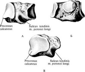
The anatomy of the foot is not as complex as in the skull. The sphenoid bone is not one here, there are three of them. They are located in front of the scaphoid. They are part of the midfoot.
Articularly connected to each other and the metatarsal bones of the foot. The intermediate cuneiform bone is somewhat shorter than the other two.
The wide side of the intermediate and lateral faces upwards, while the third bone faces downwards. The posterior articular areas form the navicular articulation. There are also articular platforms on the sides of contact with each other and at the junctions with other elements of the foot.
Bone damage
A midfoot fracture is rare. You can only get such an injury as a result of a direct blow or a fall of a heavy object.
By nature, it is more often a fracture without displacement or comminuted. Complicated by torn ligaments. Located at the inner edge of the foot, the medial bone is more susceptible to external impact, but this does not exclude the fracture of all three bones.
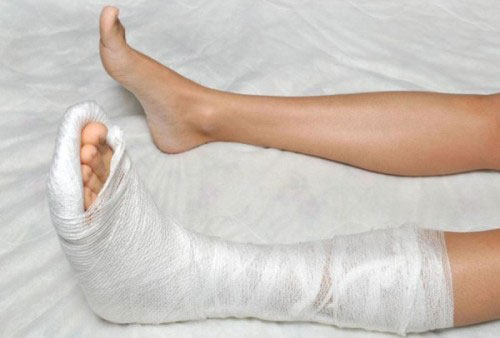
During treatment, local anesthesia, arch modeling, immobilization for 1.5 months are performed. After that, a complex of rehabilitation and preventive measures is required.
As you can see, the described elements of the skeleton, apart from the name, have little in common. But knowing the location of the bone, as well as taking into account the peculiarities of its structure, it is possible to foresee the consequences of the injuries received.
(os sphenoidale), unpaired, airy, located in the middle of the base of the skull (Fig. 1, 2). It connects with many bones of the skull and takes part in the formation of a number of bone cavities, pits, and partially in the formation of the cranial vault. 4 parts are distinguished in the bone: the body and 3 pairs of processes, of which 2 pairs are directed laterally and are called small and large wings. The third pair of processes (pterygoid) is turned downwards.
body makes up the middle part of the bone and contains the sphenoid sinus (sinus sphenoidalis), which is divided by a septum into 2 halves. The posterior surface of the body fuses with the basilar part of the occipital bone in children through cartilage, in adults through bone tissue.
Front surface the body faces the nasal cavity, adjoins the posterior cells of the ethmoid bone, closing them behind wedge-shaped shells (conchae sphenoidales). Passes along the midline of the anterior surface wedge-shaped ridge (crista sphenoidalis) on either side of which are sphenoid sinus apertures. Through them, the sinus communicates with the nasal cavity. The perpendicular plate of the ethmoid bone lies in front of the sphenoid crest. From top to bottom, the wedge-shaped ridge passes into wedge-shaped beak (rostrum sphenoidale).
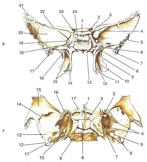
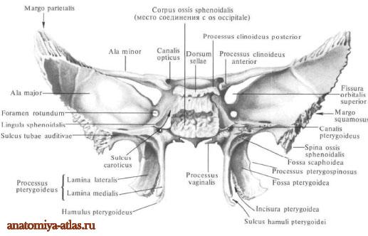
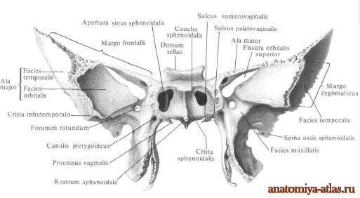
Rice. 1.
a - topography of the sphenoid bone;
b - front view: 1 - body of the sphenoid bone; 2 - wedge-shaped shell; 3 - small wing; 4 - upper orbital fissure; 5 - temporal surface of the large wing; 6 - spine of the sphenoid bone; 7 - maxillary surface; 8 - wedge-shaped ridge; 9 - pterygoid canal; 10 - round hole; 11 - infratemporal crest; 12 - orbital surface of the greater wing; 13 - aperture of the sphenoid sinus;
c - rear view: 1 - back of the Turkish saddle; 2 - pituitary fossa; 3 - anterior inclined process; 4 - upper orbital fissure; 5 - a large wing of the sphenoid bone; 6 - pterygoid canal; 7 - spine of the sphenoid bone; 8 - scaphoid fossa; 9 - lateral plate of the pterygoid process; 10 - pterygoid fossa; 11 - pterygoid notch; 12 - furrow pterygoid hook; 13 - vaginal process; 14 - pterygoid hook; 15 - pterygoid process; 16 - carotid furrow: 17 - furrow auditory tube; 18 - wedge-shaped tongue; 19 - round hole; 20 — a brain surface of a big wing; 21 - parietal edge of the large wing; 22 — small wing; 23 - visual channel; 24 - posterior surface of the body of the sphenoid bone;
d - bottom view: 1 - wedge-shaped beak; 2 - coulter; 3 - pterygoid fossa; 4 - lateral plate of the pterygoid process; 5 - oval hole; 6 - spinous opening; 7 - medial plate of the pterygoid process; 8 - opener wing; 9 - body of the sphenoid bone; 10 - scaphoid fossa; 11 - furrow of the auditory tube; 12 - spine of the sphenoid bone; 13 - infratemporal surface of the large wing; 14 - infratemporal crest; 15 - temporal surface of the large wing; 16 — small wing; 17 - wedge-shaped shells
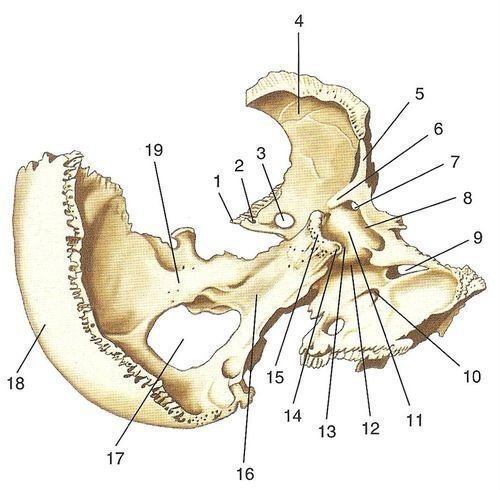
Rice. 2. Sphenoid bone and occipital bone, rear view, right and top: 1 - spine of the sphenoid bone; 2 - spinous opening; 3 - oval hole; 4 - a large wing of the sphenoid bone; 5 - small wing; 6 - anterior inclined process; 7 - visual channel; 8 - precross furrow; 9 - upper orbital fissure; 10 - round hole; 11 - tubercle of the saddle; 12 - carotid furrow; 13 - pituitary fossa; 14 - posterior inclined process; 15 - back of the saddle; 16 - slope; 17 - a large hole; 18 - occipital scales; 19 - lateral part of the occipital bone
On lateral surface bodies on each side carotid groove (sulcus caroticus) to which the internal carotid artery is adjacent. Behind and laterally, the edge of the furrow forms a protrusion - wedge-shaped tongue (lingula sphenoidalis).
Top surface body, facing the cranial cavity, forms the so-called Turkish saddle (sella turcica)(see Fig. 2). At its bottom is pituitary fossa in which the pituitary gland is located. In front and behind the fossa is bounded by protrusions, the anterior of which is represented by saddle tubercle (tuberculum sellae), and the rear - a high ridge called saddle back (dorsum sellae). The corners of the back of the Turkish saddle are extended down and back in the form posterior inclined processes (processus clinoidei posterior). On each side of the tubercle of the saddle is middle inclined process (processus clinoideus medius).
In front of the tubercle of the saddle, on wedge-shaped eminence (jugum sphenoidalis) there is a transversely running shallow precross groove (sulcus prehiasmatis), behind which is the optic chiasm.
Human Anatomy S.S. Mikhailov, A.V. Chukbar, A.G. Tsybulkin
Sphenoid bone, os sphenoidale, unpaired, forms the central section of the base.
The middle part of the sphenoid bone is the body, corpus, cubic in shape, has six surfaces. On the upper surface, facing the cranial cavity, there is a recess - the Turkish saddle, sella turcica, in the center of which is the pituitary fossa, fossa hypophysialis. It contains the pituitary gland, hypophysis. The size of the fossa depends on the size of the pituitary gland. The border of the Turkish saddle in front is the tubercle of the saddle, tuberculum sellae. Behind it, on the lateral surface of the saddle, there is an unstable middle inclined process, processus clinoideus medius.
Anterior to the tubercle of the saddle is a shallow transverse precross groove, sulcus prechiasmatis. Behind her lies the visual cross, chiasma opticum. Laterally, the groove passes into the optic canal, canalis opticus. Ahead of the furrow is a smooth surface - a wedge-shaped elevation, jugum sphenoidale, connecting the small wings of the sphenoid bone. The front crane of the upper surface of the body is serrated, protrudes slightly forward and connects with the posterior edge of the cribriform plate, forming a wedge-ethmoid suture, sutura spheno-ethmoidalis. The back border of the Turkish saddle is the back of the saddle, dorsum sellae, which ends on the right and left with a small posterior inclined process, processus clinoideus posterior.
On the sides of the saddle from back to front there is a carotid groove, sulcus caroticus (a trace and the accompanying nerve plexus). At the posterior edge of the furrow, on its outer side, a pointed process protrudes - a wedge-shaped tongue, lingula sphenoidalis.
The rear surface of the saddle back goes into upper surface basilar part, forming a slope, clivus (on it lie the bridge, medulla oblongata, basilar artery and its branches). The rear surface of the body is rough; through the cartilaginous layer, it connects to the anterior surface of the basilar part of the occipital bone and forms wedge-occipital synchondrosis, synchondrosis spheno-occipitalis. Cartilage is replaced with age bone tissue and both bones are fused.
The anterior surface of the body and part of the lower face into the nasal cavity. A wedge-shaped ridge protrudes in the middle of the anterior surface, crista sphenoidalis; its front edge is adjacent to the perpendicular plate of the ethmoid bone. The lower process of the crest is pointed, extended downwards and forms a wedge-shaped beak, rostrum sphenoidale. The latter connects with the wings, alae vomeris, forming a vomer-beak canal, canalis vomerorostratis, lying along the midline between the upper edge of the vomer and the wedge-shaped beak. Lateral to the ridge lie thin curved plates - wedge-shaped shells, conchae sphenoidales. The shells form the anterior and partly the lower walls of the sphenoid sinus, sinus sphenoidalis. Each shell has a small opening - the aperture of the sphenoid sinus, apertura sinus sphenoidalis. Outside of the aperture, there are small depressions that cover the cells of the posterior part of the labyrinth of the ethmoid bone. The outer edges of these recesses are partially connected to the orbital plate of the ethmoid bone, forming a sphenoid-ethmoid suture, sutura spheno-ethmoidalis, and the lower ones - with the orbital process, processus orbitalis, of the palatine bone.
The sphenoid sinus, sinus sphenoidalis, is a paired cavity that occupies most of the body of the sphenoid bone; it belongs to the air-bearing paranasal sinuses. The right and left sinuses are separated from one another by the septum of the sphenoid sinuses, septum sinuum sphenoidalium. which anteriorly continues into the wedge-shaped ridge. As in the frontal sinuses, the septum is often asymmetrical, as a result of which the size of the sinuses may not be the same. Through the aperture of the sphenoid sinus, each sphenoid sinus communicates with the nasal cavity. The cavity of the sphenoid sinus is lined with a mucous membrane.
Lesser wings, alae minores, of the sphenoid bone extend on both sides from the anterior superior corners of the body in the form of two horizontal plates, at the base of which there is a rounded opening. From this hole begins a bone canal up to 5-6 mm long - the visual canal, canalis opticus. It contains the optic nerve, n. opticus, and ophthalmic artery, a. ophthalmica, Small wings have an upper surface facing the cranial cavity, and a lower surface directed into the cavity of the orbit and closing the upper orbital fissure from above, fissura orbitalis superior.
The anterior margin of the lesser wing, thickened and serrated, connects to the orbital part. The posterior edge, concave and smooth, protrudes freely into the cranial cavity and is the boundary between the anterior and middle cranial fossae, fossae cranii anterior et media. Medially, the posterior edge ends with a protruding, well-defined anterior inclined process, processus clinoideus anterior (part of the dura mater is attached to it - the diaphragm of the Turkish saddle, diaphragma sellae).
Large wings, alae majores, depart from the lateral surfaces of the body of the sphenoid bone and go outward.
The large wing has five surfaces and three edges. The upper cerebral surface, facies cerebralis, is concave, facing the cranial cavity. It forms the anterior part of the middle cranial fossa. Finger-like impressions stand out on it, impressiones digitatae, and arterial grooves, sulci arteriosi (imprints of the relief of the adjacent surface of the brain and middle meningeal arteries).
There are three permanent openings at the base of the wing: a round opening, foramen rotundum, is located inward and anteriorly (the maxillary nerve, n maxillaris, exits through it); outside and behind the round hole is an oval hole, foramen ovale (it passes the mandibular nerve, n. mandibularis), and outside and behind the oval hole is a spinous hole, foramen spinosum (the middle meningeal artery, vein and nerve come through it). In addition, non-permanent holes occur in this area. One of them is the venous opening, foramen venosum, located somewhat posterior to the foramen ovale. It passes the vein going from the cavernous sinus to the pterygoid venous plexus. The second is the stony opening, foramen petrosum, through which the small stony nerve passes, the pterygofrontal suture, sutura sphenofrontalis. The outer sections of the frontal edge end with a sharp parietal edge, margo parietalis, which, with a wedge-shaped angle to the theme of another bone, forms a wedge-parietal suture, sutura sphenoparietalis. The internal sections of the frontal margin pass into a thin free margin, which is separated from the lower surface of the lesser wing, limiting the superior orbital fissure from below.
The anterior zygomatic margin, margo zygomaticus, is serrated. The frontal process, processus frontalis, the zygomatic bone and the zygomatic edge are connected, forming a sphenoid-zygomatic suture, sutura sphenozygomatica.
The posterior scaly edge, margo squamosus, connects to the wedge-shaped edge, margo sphenoidalis, and forms a wedge-scaly suture, sutura sphenosquamosa. Posteriorly and outwardly, the scaly edge ends with the spine of the sphenoid bone (the place of attachment of the sphenomandibular ligament, lig sphenomandibularis, and bundles, straining the palatine curtain, m. tensor veli palatini).
Inward from the spine of the sphenoid bone, the posterior edge of the large wing lies in front of the stony part, pars petrosa, temporal bone and limits the wedge-stony gap, fissura sphenopetrosa, medially passing into a ragged hole, foramen la-lacerum; on a non-macerated skull, this gap is filled with cartilaginous tissue and forms wedge-stony synchondrosis, synchondrosis sphenopetrosa.
Pterygoid processes, processus pterygoidei, depart from the junction of the large wings with the body of the sphenoid bone and go down. They are formed by two plates - lateral and medial. The lateral plate, lamina lateralis (processus pterygoidei), is wider, thinner and shorter than the medial one (the lateral pterygoid muscle, m. pterygoideus lateralis, starts from its outer surface).
The medial plate, lamina medialis (processus pterygoidei), is narrower, thicker and slightly longer than the lateral one. Both plates grow together with their front edges and, diverging posteriorly, limit the pterygoid fossa, fossa pterygoidea (the medial pterygoid muscle, m. pterygoideus medialis, begins here). In the lower finished
both plates do not fuse and limit the pterygoid notch, incisura pterygoidea. It contains the pyramidal process, processus pyramidalis, of the palatine bone. The free end of the medial plate ends with a pterygoid hook directed downward and outward, hamulus pterygoideus, on the outer surface of which there is a furrow of the pterygoid hook, sulcus hamuli pterygoidei (the tendon of the muscle that strains the palatine curtain, m. tensor veli palatini, is thrown through it).
The posterior superior edge of the medial plate expands at the base and forms a navicular fossa, fossa scaphoidea, of a wadded shape.
Outside of the scaphoid fossa, there is a shallow furrow of the auditory tube, sulcus tubae auditivae, which laterally passes to the lower surface of the posterior edge of the large wing and reaches the spine of the sphenoid bone (the cartilaginous part of the auditory tube is adjacent to this groove). Above the scaphoid fossa and medially there is an opening with which the pterygoid canal begins, canalis pterygoideus (vessels and nerves pass through it).
The canal runs in the sagittal direction in the thickness of the base of the pterygoid process and opens on the maxillary surface of the greater wing, on the posterior wall of the pterygopalatine fossa.
The medial plate at its base passes into a flat, horizontally directed vaginal process, processus vaginalis, which is located under the body of the sphenoid bone, covering the side of the vomer wing, ala vomeris. At the same time, the furrow of the vaginal process facing the wing of the vomer, the vomerovaginal sulcus, sulcus vomerovaginalis, turns into the vomerovaginal canal, canalis vomerovaginalis.
Outside of the process there is a sagittally running small palatovaginal groove, sulcus palatovaginalis. The sphenoid process of the palatine bone adjacent to the bottom, processus sphenoidalis ossis palatini, closes the groove into the canal of the same name, canalis palatovaginalis (the nerve branches of the pterygopalatine ganglion pass through the vomerovaginal and palatovaginal canals, and in the palatovaginal canal, in addition, the branches of the sphenoid-palatine arteries).
Sometimes, from the posterior edge of the outer plate towards the spine of the sphenoid bone, the pterygoid process, processus pterygospinosus, is directed, which can reach the indicated spine and form a hole.
The anterior surface of the pterygoid process connects to the posterior surface upper jaw in the region of the medial edge of the tubercle, forming a sphenomaxillary suture, sutura sphenomaxillaris, which lies deep in the pterygopalatine fossa.
You will be interested in this read:
Small wings (alae minores) sphenoid bone depart from the body on each side with two roots, between which is optic canal passing through the optic nerve and ophthalmic artery.
The front edges of the wings are connected to the frontal bone, the rear edge protrudes into the cranial cavity. On the back edge on each side there is anterior clinoid process. The upper surface of the small wings faces the cranial cavity, the lower surface - into the orbit and, together with the large wings, limits superior orbital fissure (fissura orbitalis superior). Pairs III, IV, VI pass through the superior orbital fissure cranial nerves, ophthalmic nerve and some other education.
Large wings (alae majores) depart from each side of the lower lateral parts of the body of the sphenoid bone. They distinguish 5 surfaces and 4 edges. Cerebral surface (fades cerebralis) facing the cranial cavity, concave, has cerebral elevations and depressions of the convolutions (finger-shaped). At the base of the large wings, 3 holes are defined on this surface: round (foramen rotundum), oval (foramen ovale) And spinous (foramen spinosum). The second branch passes through the round opening, the third branch of the trigeminal nerve passes through the oval opening, and the middle meningeal artery passes through the spinous foramen. Posteriorly, large wings end with a sharp ledge facing downwards, spine of the sphenoid bone (spina ossis sphenoidalis).
The outer surface of the large wing is divided transversely infratemporal crest (crista infratemporal) into 2 parts, of which the upper, temporal surface (fades temporalis), participates in the formation of the temporal fossa, and the lower, infratemporal surface, goes to the base of the skull and takes part in the formation of the infratemporal fossa.
Orbital surface (fades orbitalis) facing forward and forms the posterior part of the lateral wall of the orbit. facing the upper jaw maxillary surface, on which a round hole opens from the side of the pterygopalatine fossa.
The edges of the large wings are connected to the squamous part of the temporal bone, with the zygomatic, parietal and frontal bones. The name of the regions corresponds to the adjacent bones: margo squamosus, margo zygomaticus, margo parietalis and margo frontalis.
pterygoid processes(processus pterygoideus) composed of medial and lateral plates (laminae medialis et lateralis). In front, both plates are connected, and behind they are separated from each other by a deep pterygoid fossa (fossa pterygoidea). At the bottom between both plates there is pterygoid notch (incisura pterygoidea), which includes the pyramidal process of the palatine bone. The medial plate below ends in a curved pterygoid hook (hamulus pterygoideus), through which the tendon of the muscle that strains the palatine curtain is thrown. In the upper part of the posterior edge of the medial plate is navicular fossa (fossa scaphoidea). From it begins a muscle that strains the palatine curtain. The cartilaginous part of the auditory tube is adjacent to the upper part of the medial plate of the pterygoid process.
Passes along the anterior margin of the pterygoid processes greater palatine sulcus (sulcus palatinus major). At the base of the pterygoid process in the anteroposterior direction is located pterygoid canal (canalis pterygoideus). In 10% of cases, a pterygo-spinous plate is formed between the lateral plate of the pterygoid process and the spine of the sphenoid bone, which covers the foramen ovale and prevents anesthesia from being carried out near it.
Ossification: the development of most of the bone occurs on the basis of cartilage, a smaller part (the medial plate of the pterygoid processes and the lateral sections of the large wings) develops due to the connective tissue. Ossification points arise in the anterior and posterior parts of the body, in each of the processes, and individual ossification points appear in the medial plate of the pterygoid processes. The ossification points in the large wings appear first in the 2nd month of embryo development, and all the rest appear in the 3rd month. At the 6-7th month of the intrauterine period, the small wings are connected to the anterior half of the body of the sphenoid bone, and by the end of this period, the anterior and posterior parts of the body merge. Large wings and sphenoid processes are connected to the body of the bone at the end of the 1st year of life. The sphenoid sinus begins to form in the 3rd year of life and reaches full development by 12-15 years. The connection of the body of the sphenoid bone with the basilar part of the occipital bone occurs in the period from 16 to 20 years, more often at 16-18 years.
Human Anatomy S.S. Mikhailov, A.V. Chukbar, A.G. Tsybulkin
Body of the sphenoid bone corpus ossis sphenoidalis, the middle part of the bone, cubic in shape, has six surfaces. The upper surface of the body, facing the cranial cavity, has a recess in its middle sections - the Turkish saddle, sella turcica. in the center of which is the pituitary fossa. It contains the pituitary gland. The size of the fossa is determined by the size of the pituitary gland. The pituitary fossa is especially vulnerable in case of premature birth. The fusion of the two nuclei of ossification of the fossa occurs on the 8th month of intrauterine life. Hence, there is a possibility of damage to the structure of the pituitary fossa with subsequent dysfunction of the pituitary gland. The Turkish saddle is limited in front by the tubercle of the saddle, tuberculum sellae. Behind it, on the lateral surface of the saddle, there is a non-permanent middle inclined process, processus clinoideus medius. Anterior to the tubercle of the saddle there is a shallow transverse furrow of the decussation, sulcus chiasmatis. On it lies the optic chiasm, chiasma opticum. On the sides, the furrow passes into the optic canal, canalis opticus. Ahead of the furrow is a smooth surface - a wedge-shaped elevation, jugum sphenoidale connecting the small wings of the sphenoid bone. The anterior edge of the upper surface of the body is serrated, protrudes slightly forward and connects with the posterior edge of the perforated plate, lamina cribrosa, ethmoid bone, forming a wedge-ethmoid suture, sutura sphenoethmoidalis. The perforated plate has a large number of holes (25-30), through which branches of the anterior ethmoid (olfactory) nerve and the vein accompanying the anterior ethmoid artery pass from the nasal cavity into the cranial cavity (there are olfactory grooves on the sides of the anterior edge of the sphenoid bone). If the sense of smell is impaired or absent, the kinetics of the anterior edge of the sphenoid bone should be checked. As a result of trauma to the frontal bone, there may be a violation of the ratio in the wedge-lattice suture, followed by traumatization of the olfactory bulbs.
The Turkish saddle is bounded at the back by the back of the saddle, dorsum sellae, which ends on each side with a small posterior inclined process, processus clinoideus posterior. On the sides of the Turkish saddle, from back to front, there is a carotid furrow, sulcus caroticus(the imprint of the internal carotid artery and accompanying nerve plexus).
Rice. Sphenoid bone (according to H. Feneis, 1994): 1 - body; 2 - wedge-shaped elevation; 3 - large wing, 4 - small wing; 5 - precross furrow; 6 - Turkish saddle; 7 - pituitary fossa; 8 - anterior inclined process; 9 - posterior inclined process; 10 - back of the saddle; 11 - carotid groove; 12 - wedge-shaped ridge; 13 - wedge-shaped beak; 14 - aperture of the sphenoid sinus; 15 - visual channel; 16 - superior orbital fissure; 17 - cerebral surface; 18 - temporal surface; 19 - orbital surface; 20 - zygomatic edge; 21 - frontal edge; 22 - parietal edge; 23 - scaly edge; 24 - infratemporal crest; 25 - round hole; 26 - oval hole; 27 - spinous opening; 28 - spine of the sphenoid bone; 29 - pterygoid (Vidian) canal; 30 - pterygoid process; 31 - lateral plate of the pterygoid process; 32 - medial plate of the pterygoid process; 33 - pterygoid hook; 34 - pterygoid notch; 35- wedge-shaped surface sphenobasilar synchondrosis.
The back surface of the back of the saddle passes into the upper surface of the basilar part of the occipital bone, forming a slope, clivus. On the slope are the bridge, the medulla oblongata, the basilar artery with its branches. The posterior surface of the body is rough. Through the cartilaginous layer, it connects to the anterior surface of the basilar part of the occipital bone, forming the sphenoid-occipital synchondrosis (SSO), Synchondrosis sphenooccipitalis. More often in the osteopathic literature and among osteopaths, another term is found - sphenobasilar symphysis. Despite the existence of the International Nomenclature, the last anatomical term has taken root and is most common among osteopaths. It is believed that by the age of 25, cartilage is replaced by bone tissue and both bones fuse. However, there is still no consensus on this issue. Probably, the bones are still not fully fused.
The front and part of the lower surface of the body face the nasal cavity. In the middle of the anterior surface of the body, a vertically running wedge-shaped ridge protrudes, Crista sphenoidalis. Its anterior edge is adjacent to the posterior edge of the perpendicular plate, lamina perpendicularis, ethmoid bone. The lower segment of the crest is pointed, extended downwards, and forms a wedge-shaped beak, rostrum sphenoidale, which is wedged between the opener wings, alae vomeris. On the sides of the ridge lies a thin curved plate - a wedge-shaped shell, concha sphenoidalis. This shell, forming the anterior and partly inferior walls of the sphenoid sinus, sinus sphenoidalis, has a small opening - the aperture of the sphenoid sinus, apertura sinus sphenoidalis. Outside of the aperture there are small depressions that cover the cells of the posterior part of the labyrinth of the ethmoid bone. The outer edges of these recesses are partially connected to the orbital plate of the ethmoid bone, forming a sphenoid-ethmoid suture, sutura sphenoethmoidalis, and the lower ones - with the orbital process, processus orbitalis, palatine bone.
sphenoid sinus, sinus sphenoidalis, a steam cavity, performs most of the body of the sphenoid bone and belongs to the air-bearing paranasal sinuses. Both right and left sinuses are separated from one another by the septum of the sphenoid sinuses, which continues anteriorly into the sphenoid crest. As in the frontal sinuses, the septum sometimes lies asymmetrically, as a result of which the size of both sinuses may not be the same. Through the aperture, the cavity of each sphenoid sinus opens into nasal cavity. The cavity of the sphenoid sinus is lined with a mucous membrane.
small wings, alae minores, sphenoid bone with two roots depart in both directions from the anterior-upper corners of the body in the form of two horizontally located plates, at the base of which there is a rounded hole. It represents the beginning of the bone canal up to 5-6 mm long - the visual canal, canalis opticus. It contains the optic nerve n. opticus, and the ophthalmic artery, a. ophthalmica. Small wings have an upper surface facing the cranial cavity, and a lower surface directed into the cavity of the orbit and closing the upper orbital fissure from above, fissura orbitalis superior. The anterior margin of the lesser wing, thickened and serrated, is connected to the orbital part of the frontal bone. The posterior concave and smooth edge protrudes freely into the cranial cavity and is the boundary between the anterior and middle cranial fossae, fossae cranii anterior et media. Medially, the posterior edge ends with a protruding, well-defined, anterior inclined process, processus clinoideus anterior(part of the solid is attached to it meninges, which forms the diaphragm of the Turkish saddle, diaphragma sellae).
Large wings of the sphenoid bone, alae majores, depart from the lateral surfaces of the body of the sphenoid bone and are oriented outwards. The large wing has five surfaces and three edges. superior cerebral surface, facies cerebralis, concave and turned into the cranial cavity. It forms the anterior part of the middle cranial fossa and bears sulcular depressions, cerebral eminences and arterial sulci, sulci arteriosi(imprints of the relief of the adjacent surface of the brain and middle meningeal arteries). There are three holes at the base of the large wing: a round hole is located inward and anteriorly, foramen rotundum(the maxillary nerve exits through it, n. maxillaris). Outside and behind the round is an oval hole, foramen ovale (it passes the mandibular nerve, n. mandibularis, and the vasculature of the foramen ovale). Still outside and posterior to the foramen ovale is the spinous foramen, foramen spinosum(through it pass the middle meningeal artery, vein and nerve). Antero-superior, orbital surface, facies orbitalis, smooth, diamond-shaped, turned into the cavity of the orbit, where it forms most of its outer wall. The lower edge of this surface is separated from the posterior edge of the orbital surface of the body of the upper jaw; here the inferior orbital fissure is formed, fissura orbitalis inferior. Anterior, maxillary surface, facies maxillaris, a small area of triangular shape, bounded above by the orbital surface, and from the side and below by the root of the pterygoid process of the sphenoid bone. It is part of the posterior wall of the pterygopalatine fossa, fossa pterygopalatina. There is a round hole on the surface. Upper lateral, temporal surface, facies temporalis, somewhat concave, takes part in the formation of the wall of the temporal fossa, fossa temporalis(the temporalis muscle is attached to it, m. temporalis). From below, this surface is bounded by the infratemporal crest, crista infratemporalis, below which the surface is located, where the oval hole opens, foramen ovale, and a spinous foramen. It forms the superior wall of the infratemporal fossa fossa infratemporalis. Here begins part of the lateral pterygoid muscle, m. pterygoideus lateralis. The upper, frontal, edge is widely serrated, connects with the orbital part of the frontal bone in the sphenoid-frontal suture ( sutura sphenofrontalis). The outer sections of the frontal edge end with a sharp parietal edge, margo parietalis, which with the wedge-shaped angle of the parietal bone forms a wedge-parietal suture ( sutura sphenoparietalis). The internal sections of the frontal margin pass into a thin free margin, which is separated from the lower surface of the lesser wing, limiting the upper orbital fissure from below fissura orbitalis superior. Anterior, zygomatic edge, margo zygomaticus, serrated, connects with the frontal process, processus frontalis, zygomatic bone, forming a wedge-zygomatic suture ( sutura sphenozygomatica). Back, scaly edge, margo squamosus, connects to the wedge-shaped edge, margo sphenoidalis, temporal bone in the sphenoid-squamous suture ( sutura sphenosquamosa). Posteriorly and outwards, the scaly edge ends with the spine of the sphenoid bone, spina ossis sphenoidalis. Here is the site of attachment of the sphenomandibular ligament, lig. sphenomandibular, and bundles of muscles that strain the palatine curtain, m. tensor veli palatini. Inward from the spine of the sphenoid bone, the posterior edge of the large wing lies in front of the petrous part, pars petrosa, temporal bone and limits the sphenoid-stony fissure, fissura sphenopetrosa, medially passing into a torn hole, foramen lacerum. This gap is filled with cartilaginous tissue, forming wedge-stony synchondrosis, synchondrosis sphenopetrosa.
pterygoid processes, processus pterygoidei, depart from the junction of the large wings with the body of the sphenoid bone and go down. The pterygoid processes are formed by two plates - lateral and medial. lateral plate, lamina lateralis processus pterygoidei, wider, but thinner and shorter than the inner one (the lateral pterygoid muscle begins from its outer surface, m. pterygoideus lateralis). medial plate, lamina medialis processus pterygoidei, narrower, thicker and slightly longer than the outer. Both plates grow together with their front edges and, diverging posteriorly, limit the pterygoid fossa, fossa pterygoidea(here begins the medial pterygoid muscle, m. pterygoideus medialis). In the lower sections, both plates do not fuse and limit the pterygoid notch, incisura pterygoidea filled with pyramidal process, processus pyramidalis, palatine bone. The free end of the inner plate ends with a pterygoid hook directed downward and outward, hamulus pterygoideus, on the outer surface of which there is a furrow of the pterygoid hook, sulcus hamuli pterygoidei(through it the tendon of the muscle straining the palatine curtain is thrown, m. tensor veli palatini). The posterior-upper edge of the inner plate at the base expands and forms an oblong navicular fossa, fossa scaphoidea(bunches of muscles begin in it, straining the palatine curtain, m. tensor veli palatini). Outside of the scaphoid fossa is a shallow furrow of the auditory tube, sulcus tubae audilivae, which laterally passes to the large wing and reaches the spine of the sphenoid bone (the cartilaginous part of the auditory tube is adjacent to this groove). Above the scaphoid fossa and medial from it there is an opening leading to the pterygoid canal, canalis pterygoideus(vessels and nerves pass through it). The canal runs in the sagittal direction in the thickness of the base of the pterygoid process and opens on the maxillary surface of the greater wing of the sphenoid bone on the posterior wall of the pterygopalatine fossa. Under the outlet, along the anterior face of the pterygoid process, there is a pterygopalatine groove. The inner plate at its base gives off a flat, horizontally running vaginal process directed inwards, processus vaginalis, which is located under the body of the sphenoid bone, covering the wing of the vomer from the side. As a result of this, the groove of the vaginal process facing the wing is the vomerovaginal groove, sulcus vomerovaginalis, turns into the vomerovaginal canal, canalis vomerovaginalis. Outside of the process, there is sometimes a sagittally running small palatovaginal groove, Sulcus palatovaginalis. In the latter case, the sphenoid process of the palatine bone adjacent from below closes the groove into the canal of the same name (the nerve branches of the pterygopalatine ganglion pass through both canals, and the branches of the sphenoid-palatine artery also pass through the palatovaginal canal). Sometimes, from the posterior edge of the outer plate, the pterygoid process is directed towards the spine of the sphenoid bone. processus pterygospinosus, which can reach the specified awn and form a hole.