Musculoskeletal system organism
The skeleton (from the Greek skeletos - dried up, dried) is a complex of bones that perform many functions: supporting, protective, locomotor, shaping, overcoming gravity. The total mass of the skeleton is from "/7 to "/5 of the human body weight. The human skeleton consists of more than 200 bones, 33-34 bones of the skeleton are not paired. These are the vertebrae, sacrum, coccyx, some bones of the skull and sternum, the rest of the bones are paired. The skeleton is conditionally divided into two parts: axial and additional. To axial skeleton includes the spinal column (26 bones), skull (29 bones), chest (25 bones); to the additional - the bones of the upper (64) and lower (62) limbs.The bones of the skeleton are levers driven by muscles. As a result, body parts change position relative to each other and move the body in space. Ligaments, muscles, tendons, fascia are attached to the bones, which are elements of the soft skeleton or soft skeleton, which also takes part in holding organs near the bones that form a hard (hard) skeleton. The skeleton forms a container for organs, protecting them from external influences: the brain is located in the cranial cavity, the spinal cord is in the spinal canal, the heart, large vessels, lungs, esophagus, etc. are in the chest, and the genitourinary organs are in the pelvic cavity.
Bones are an unusually complex and very strong complex of spatial systems, which prompted architects to create "perforated structures".
Bones can withstand heavy loads. So, the tibia can withstand a weight that is 2 thousand times its weight (1650 kg), brachial bone- 850 kg, tibia - up to 1500 kg.
Bones are involved in mineral metabolism, they are a depot of calcium, phosphorus, etc. Living bone contains vitamins L, D, C, etc. The vital activity of the bone depends on the functions of the pituitary gland, thyroid and parathyroid glands, adrenal glands and sex glands (gonads).
The skeleton is formed by varieties of connective tissue - bone and cartilage, which consist of cells and dense intercellular substance. Bones and cartilage are closely related to each other by a common structure, origin and function. Most bones (bones of the limbs, base of the skull, vertebrae) develop from cartilage, their growth is ensured by proliferation (an increase in the number of cells). A small number of bones develop without the participation of cartilage (bones of the roof of the skull, lower jaw, clavicle).
Some cartilages are not connected with the bone and do not change throughout a person's life (cartilages of the auricles, airways). Some cartilages are functionally related to the bone (articular cartilages, menisci).
In the human embryo and other vertebrates, the cartilaginous skeleton makes up about 50% of the total body mass. However, cartilage is gradually replaced by bone, in an adult, the mass of cartilage reaches about 2% of body weight. These are articular cartilages, intervertebral discs, cartilages of the nose and ear, larynx, trachea, bronchi and ribs. Cartilages perform the following functions:
1) cover the articulated surfaces, which are therefore highly resistant to wear;
2) articular cartilage and intervertebral discs, which are the objects of application of compression and tension forces, carry out their transmission and shock absorption;
3) the cartilages of the airways and the outer ear form the walls of the cavities. Muscles, ligaments, tendons are attached to other cartilages.
Cartilaginous tissue contains about 70-80% water, 10-15% organic matter, 4-7% salts. About 50-70% of the dry matter of cartilage is collagen. Depending on the composition of the cartilage are hyaline, elastic and collagen fibers. Like other types of connective tissue, cartilage tissue consists of a few cells (chondrocytes) and a dense intercellular substance produced by them. Cartilages do not have blood vessels; they are nourished by diffusion from the surrounding tissues.
Hyaline cartilage is smooth, shiny, bluish-white in color. It formed mainly the skeleton of the embryo, in an adult - costal cartilages, most of the cartilages of the larynx, cartilages of the nose, trachea, bronchi and articular cartilages (hyaline cartilage calcifies with age).
Elastic cartilage is less transparent, yellowish in color. Made up of elastic cartilage Auricle, vocal processes of the arytenoid cartilages of the larynx and the auditory tube.
Fibrous cartilage forms intervertebral discs, menisci of the knee and temporomandibular joints. Fibrous cartilage is found in the areas of attachment of ligaments and tendons to bones and cartilage.
Bones are formed by bone tissue, the mechanical properties of which determine the functions of the bones. Thus, the tensile strength of fresh bone and pure copper is the same and 9 times greater than the resistance of lead. Bone withstands a compression of 10 kg/mm2 (similar to cast iron), while brick only 0.5 kg/mm2. The fracture strength of the ribs is 110 kg/cm2. This is due to the peculiarities of the chemical composition, structure and architectonics of bones. The water content in the bone reaches 50%. In the dry matter bone tissue contains about 33% organic and 6-7% inorganic substances.
The bone consists of cells (osteoblasts and osteocytes) and intercellular substance. Osteoblasts are polygonal, cubic, process young cells, osteocytes are mature multi-pronged spindle-shaped cells. Osteoblasts synthesize the components of the intercellular substance and release them from the cell through the entire surface in different directions, which leads to the formation of gaps (spaces) in which they lie, turning into osteocytes.
There are two types of bone tissue: reticulofibrous (coarse-fibrous) and lamellar. Reticulofibrous bone tissue is located in the areas of attachment of the tendons to the bones, in the sutures of the skull after their overgrowth. It consists of thick disordered bundles of collagen fibers, between which there is an amorphous substance. Osteocytes lie in the lacunae.
Lamellar bone tissue is the most common in the body. It is formed by bone plates with a thickness of 4 to 15 microns, which consist of osteocytes and fine-fibrous bone ground substance. The fibers that form the plates lie parallel to each other and are oriented in a certain direction. At the same time, the fibers of neighboring plates are multidirectional and intersect almost at a right angle, which ensures greater bone strength.
The bone outside, except for the articulated surfaces, is covered with a periosteum, which is a strong connective tissue plate rich in blood and lymphatic vessels, nerves. The periosteum is firmly fused with the bone with the help of connective tissue perforating fibers that penetrate deep into the bone. In the inner layer of the periosteum there are thin spindle-shaped "resting" osteogenic cells, due to which the development, growth in thickness and regeneration of bones after damage occurs.
The bones of a living person are a dynamic structure in which there is a constant metabolism, anabolic and catabolic processes, the destruction of old and the creation of new bone plates. The bones adapt to the changing conditions of the vital activity of the organism, under the influence of which the restructuring of their macro- and microscopic structure occurs. The external shape of the bones changes under the influence of stretching and pressure, and the bones develop the better, the more intense the activity of the muscles associated with them.
vertebral column It is made up of 33 individual vertebrae. Distinguish cervical region(7 cervical vertebrae), thoracic (12 thoracic), lumbar (5 lumbar), sacral (5 sacral) and coccygeal (4 or 5 coccygeal vertebrae). The sacral and coccygeal vertebrae fuse together to form the sacrum and coccyx.
A typical vertebra has a body, a neural arch that surrounds and protects the spinal cord, and seven processes. The unpaired, backward-facing process is called the spinous process. It serves to attach ligaments and muscles. The bodies of the vertebrae are interconnected by means of intervertebral cartilages, which, together with the ligaments and muscles that run along the spine, hold the body in an upright position.
All vertebrae differ in shape and size, the first two are especially different from the others. cervical vertebrae- atlas and epistropheus. The movable connection of these vertebrae facilitates the movement of the head. The rest of the vertebrae, the lower they are, the more massive, as they experience greater heaviness. Inside the spinal column, in the spinal canal, formed by holes in the vertebrae, is the spinal cord. It is securely protected from all sides.
The spinal column has bends forward - lordosis, back (posteriorly) - kephosis, to the sides - scoliosis. The bends of the spinal column increase its spring properties, i.e. contribute to the springy movements of the spinal column. Under the influence of external influences, the bends can change during the day. Therefore, the height of the spine, and hence the height of a person, can fluctuate during the day on average from 1 to 2-2.5 cm.
The spinal column of a newborn does not have bends, they appear in the process of growth of the body. In the beginning, the newborn develops cervical lordosis (as the child begins to hold his head), then thoracic kephosis (the child begins to sit), and then lumbar lordosis(he starts to stand) and sacral kephosis. By the age of five or six, the curves are clearly visible. In children school age scoliosis can often be observed.
The thorax is back supported by the spine. On both sides of it, flat bones extend - ribs, representing curved bone plates. In the rib, a middle part (body) and two ends (anterior and posterior) are distinguished. The posterior end of the rib has a thickening - the head, which, through a composite surface, articulates with the body of the spine. Behind the head of the rib is the middle part - the neck, and behind it is the tubercle.
Each rib articulates with two vertebrae at the same time. The exceptions are the 9th (not always), 10th, 11th, 12th thoracic vertebrae, each of which is connected to one rib. The anterior ends of the ribs are directed towards the sternum. The cartilages of the upper seven pairs of ribs adhere to the sternum (true, or thoracic, ribs). The next three pairs of ribs (8, 9, 10) grow each with their own cartilage to the cartilage of the overlying pair, forming a costal arch. These are the so-called false ribs. The last two pairs (11th, 12th) do not reach the sternum and are very variable in length (free ribs).
The respiratory muscles and the diaphragm are attached to the ribs. When inhaling, the ribs are removed by the front ends from the spine forward and rise upward.
The shoulder girdle consists of two pairs of bones - the shoulder blades and the clavicles. Bones and joints shoulder girdle give the hand support and firmly connect it to the body.
The pelvic girdle is formed by three pairs of bones: ischial, pubic and iliac. The bones of the pelvis support the entire weight of the body.
The skeleton of the upper limbs is formed by: the humerus, the radius and ulna of the forearm, eight small bones of the wrist, five thin metacarpal bones and phalanges of the fingers. Each finger has three phalanges, except for the thumb, which has only two.
The skeleton of the lower extremities consists of the femur (thigh), tibia and fibula (in the lower leg), 7 tarsal bones (in the ankles and heel), 5 metatarsal bones (in the forefoot) and 14 phalanges of the fingers.
The skull has two sections: cerebral and facial. The brain skull protects the brain. The bone plates of which it consists are distinguished by great strength. The cranial box is formed by the following bones: frontal, two temporal, occipital, two maxillary, two zygomatic, two nasal, vomer, two lacrimal, hyoid bone, palatine. The only movable bone of the skull is the lower jaw.
Some bones of the skull are pierced by sinuses containing air (maxillary, frontal, sinuses of the main and ethmoid bones). This reduces the overall weight of the skull. It is connected to the spine by two occipital condyles.
Abstract on biology on the topic:
Pupil 9 "G" class
high school № 117
SWAD Moscow
Yuditsky Alexander.
Moscow 2004
Plan:
I. Introduction.
II. Skeleton.
1. Spine.
2. Chest.
3. Limbs.
4. Leg and arm.
III. Two kinds muscle tissue.
1.Smooth muscles.
2. Muscles of the skeleton.
3. Nerve connections in the muscles.
4. Muscles generate heat.
5. Strength and speed of muscle contraction.
IV. Tiredness and rest.
1. Causes of fatigue.
v. Statics and dynamics of the human body.
1. Conditions of equilibrium.
VI. Everyone needs sports.
1. Muscle training.
2. Labor and sport.
3. Anyone can become an athlete.
VII.
VIII. Conclusion.
XI.
Musculoskeletal system
The musculoskeletal system consists of bones of the skeleton with joints, ligaments and muscles with tendons, which, along with movements, provide the supporting function of the body. Bones and joints participate in movement passively, obeying the action of muscles, but play a leading role in the implementation support function. A certain shape and structure of the bones give them greater strength, the reserve of which for compression, expansion, bending significantly exceeds the loads possible during everyday work of the support- locomotive system. For example, the human tibia can withstand a load of more than a ton during compression, and in terms of tensile strength it is almost as good as cast iron. Ligaments and cartilage also have a large margin of safety.
The skeleton is made up of interconnected bones. It provides our body with support and shape retention, and also protects the internal organs. An adult human skeleton consists of about 200 bones. Each bone has a certain shape, size and occupies a certain position in the skeleton. Part of the bones are interconnected by movable joints. They are driven by muscles attached to them.
Spine. The original structure that makes up the main support of the skeleton is the spine. If it consisted of a solid bone rod, then our movements would be constrained, devoid of flexibility and would deliver the same unpleasant sensations as riding a cart without springs on a cobblestone pavement.
The elasticity of hundreds of ligaments, cartilage layers and bends makes the spine a strong and flexible support. Thanks to this structure of the spine, a person can bend down, jump, somersault, run. Very strong intervertebral ligaments allow the most complex movements and at the same time create reliable protection for the spinal cord. It is not subjected to any mechanical stretching, pressure under the most incredible curves of the spine.
The bends of the spinal column correspond to the influence of the load on the axis of the skeleton. Therefore, the lower, more massive part becomes a support when moving; the upper, with free movement, helps to maintain balance. The vertebral column could be called the vertebral spring.
Wavy curves of the spine provide its elasticity. They appear with the development of the motor abilities of the child, when he begins to hold his head, stand, walk.
Rib cage. The thorax is formed by the thoracic vertebrae, twelve pairs of ribs and a flat sternum, or breastbone. The ribs are flat curved bones. Their rear ends are movably connected to the thoracic vertebrae, and the front ends of the ten upper ribs are connected to the sternum with the help of flexible cartilage. It provides mobility chest when breathing. The two lower pairs of ribs are shorter than the rest and terminate freely. The chest protects the heart and lungs, as well as the liver and stomach.
It is interesting to note that the ossification of the chest occurs later than other bones. By the age of twenty, the ossification of the ribs ends, and only by the age of thirty does the complete fusion of the parts of the sternum, consisting of the handle, the body of the sternum and the xiphoid process, occur.
The shape of the chest changes with age. In a newborn, it has, as a rule, the shape of a cone with the base turned down. Then the circumference of the chest in the first three years increases faster than the length of the body. Gradually, the cone-shaped chest acquires a rounded shape characteristic of a person. Its diameter is greater than its length.
The development of the chest depends on the lifestyle of a person. Compare an athlete, swimmer, athlete with a non-athlete. It is easy to understand that the development of the chest, its mobility depends on the development of the muscles. Therefore, in adolescents of twelve to fifteen years old who go in for sports, the circumference of the chest is seven to eight centimeters larger than that of their peers who do not go in for sports.
Improper seating of students at a desk, squeezing the chest can lead to its deformation, which disrupts the development of the heart, large vessels and lungs.
Limbs. Due to the fact that the limbs are attached to a reliable support, they have mobility in all directions and are able to withstand heavy physical exertion.
Light bones - clavicles and shoulder blades, lying on the upper part of the chest, cover it like a belt. This is the handhold. The protrusions and ridges on the collarbone and shoulder blade are the site of muscle attachment. The greater the strength of these muscles, the more developed the bone processes and irregularities. In an athlete, a loader, the longitudinal ridge of the scapula is more developed than in a watchmaker or accountant. The clavicle is the bridge between the bones of the trunk and arms. The shoulder blade and collarbone create a reliable spring support for the hand.
The position of the shoulder blades and collarbones can be used to judge the position of the hands. Anatomists helped to restore the broken off hands of the ancient Greek statue of Venus de Milo, determining their position by the silhouettes of the shoulder blades and collarbones.
The pelvic bones are thick, wide and almost completely fused. In humans, the pelvis justifies its name - it, like a bowl, supports the internal organs from below. This is one of the typical features of the human skeleton. The massiveness of the pelvis is proportional to the massiveness of the bones of the legs, which bear the main load when a person moves, therefore the human pelvic skeleton can withstand a large load.
Leg and hand. With a vertical posture, a person’s hands do not carry a constant load as supports, they acquire lightness and variety of action, freedom of movement. The hand can perform hundreds of thousands of different motor operations. The legs carry the entire weight of the body. They are massive, have extremely strong bones and ligaments.
Shoulder head is not limited in wide circular motions hands, For example, when throwing a spear. The head of the femur protrudes deeply into the deepening of the pelvis, which limits movement. The ligaments of this joint are the strongest and hold the weight of the body on the hips.
Exercise and training achieve greater freedom of movement of the legs, despite their massiveness. A convincing example of this can be ballet art, gymnastics, martial arts.
The tubular bones of the arms and legs have a huge margin of safety. Interestingly, the arrangement of the openwork crossbars of the Eiffel Tower corresponds to the structure of the spongy substance of the heads. tubular bones, as if J. Eiffel designed the bones. The engineer used the same laws of construction that determine the structure of the bone, giving it lightness and strength. This is the reason for the similarity of the metal structure and the living bone structure.
The elbow joint provides complex and diverse movements of the hand in the working life of a person. Only he has the ability to rotate the forearm around its axis, with a characteristic movement of unwinding or twisting.
The knee joint directs the lower leg when walking, running, jumping. knee ligaments in humans, they determine the strength of the support when the limb is straightened.
The hand begins with a group of bones of the wrist. These bones do not experience strong pressure, perform a similar function, so they are small, monotonous, and difficult to distinguish. It is interesting to mention that the great anatomist Andrei Vesalius could, blindfolded, identify each carpal bone and tell whether it belongs to the left or right hand.
The bones of the metacarpus are moderately mobile, they are located in the form of a fan and serve as a support for the fingers. Phalanges of fingers - 14. All fingers have three bones, except for the thumb - it has two bones. A person has a very mobile thumb. It can become at right angles to everyone else. His metacarpal able to oppose the rest of the bones of the hand.
Development thumb associated with labor movements of the hand. The Indians call the thumb "mother", the Javanese - "big brother". In ancient times, captives were cut off the thumb to humiliate their human dignity and make them unfit for participation in battles.
The brush makes the most subtle movements. In any working position of the hand, the hand retains complete freedom of movement.
The foot became more massive due to walking. The tarsal bones are very large and strong compared to the carpal bones. The largest of them are ram and calcaneus. They can withstand significant weight of the body. In newborns, the movements of the foot and thumb are similar to those of monkeys. Strengthening the supporting role of the foot during walking led to the formation of its arch. When walking, standing, you can easily feel how the entire space between these points “hangs in the air”.
The vault, as is known in mechanics, withstands greater pressure than the platform. The arch of the foot provides the elasticity of the gait, eliminates pressure on the nerves and blood vessels. His education in the history of the origin of man is connected with upright walking and is distinctive feature a person acquired in the process of his historical development.
Two types of muscle tissue.
Smooth muscles. When we talked about muscles, we usually thought of skeletal muscles. But, besides them, in our body in the connective tissue there are smooth muscles in the form of single cells, in some places they are collected in bundles.
Many smooth muscles in the skin, they are located at the base of the hair bag. By contracting, these muscles raise the hair and squeeze out fat from the sebaceous gland.
In the eye around the pupil are smooth circular and radial muscles. They work all the time, imperceptibly for us, work: in bright light, the circular muscles constrict the pupil, and in the dark, the radial muscles contract and the pupil expands.
In the walls of all tubular organs - the respiratory tract, blood vessels, digestive tract, urethra, etc. - there is a layer of smooth muscles. Under the influence of nerve impulses, it is reduced. For example, reducing it in the windpipe delays the flow of air containing harmful impurities - dust, gases.
Due to the contraction and relaxation of the smooth cells of the walls of blood vessels, their lumen either narrows or expands, which contributes to the distribution of blood in the body. The smooth muscles of the esophagus, contracting, push a lump of food or a sip of water into the stomach.
Complex plexuses of smooth muscle cells are formed in organs with a wide cavity - in the stomach, bladder, uterus. The contraction of these cells causes compression and narrowing of the lumen of the organ. The strength of each cell contraction is negligible, since they are very small. However, the addition of the forces of entire beams can create a contraction of enormous force. Powerful contractions create a sensation of intense pain.
Muscles of the skeleton. Skeletal muscles carry out both static activity, fixing the body in a certain position, and dynamic, ensuring the movement of the body in space and its individual parts relative to each other. Both types of muscular activity closely interact, complementing each other: static activity provides a natural background for dynamic activity. As a rule, the position of the joint is changed with the help of several muscles of multidirectional, including opposite action. Complex joint movements are performed by coordinated, simultaneous or sequential contraction of non-directional muscles. Consistency (coordination) is especially necessary for the performance of motor acts in which many joints participate (for example, skiing, swimming).
Skeletal muscles are not only the executive motor apparatus, but also a kind of sensory organs. In the muscle fiber and tendons there are nerve endings - receptors that send impulses to cells of various levels of the central nervous system. As a result, a closed cycle is created: impulses from various formations of the central nervous system, going along the motor nerves, cause muscle contraction, and impulses sent by muscle receptors inform the central nervous system about each element of the system. The cyclic system of connections ensures the accuracy of movements and their coordination. Although the movement of skeletal muscles is controlled by various sections of the central nervous system, the leading role in ensuring interaction and setting the goal of a motor reaction belongs to the cerebral cortex. In the cerebral cortex, the motor and sensory zones of the representations form a single system, with each muscle group corresponding to a certain section of these zones. Such a relationship allows you to perform movements, attributing them to factors acting on the body. environment. Schematically, the control of arbitrary movements can be represented as follows. The tasks and purpose of a motor action are formed by thinking, which determines the direction of attention and efforts of a person. Thinking and emotions accumulate and direct these efforts. The mechanisms of higher nervous activity form the interaction of psychophysiological mechanisms of movement control at various levels. Based on the interaction of the musculoskeletal system, deployment and correction of motor activity are provided. Analyzers play an important role in the implementation of the motor reaction. Motor analyzer provides dynamics and interconnection muscle contractions, participates in the spatial and temporal organization of the motor act. The balance analyzer, or vestibular analyzer, interacts with the motor analyzer when the position of the body in space changes. Vision and hearing, actively perceiving information from the environment, are involved in spatial orientation and correction of motor reactions.
The name "muscle" comes from the word "musculis", which means "mouse".
This is due to the fact that anatomists, observing the contraction of skeletal muscles, noticed that they seem to run under the skin, like mice.
A muscle is made up of muscle plexuses. The length of muscle plexuses in humans reaches 12 cm. Each such plexus forms a separate muscle fiber.
Numerous rod-shaped nuclei are located under the shell of the muscle fiber. Along the entire length of the cell stretches several hundred of the thinnest filaments of the cytoplasm - myofibrils, capable of contracting. In turn, myofibrils are formed by 2.5 thousand protein filaments.
In myofibrils, light and dark discs alternate, and under a microscope, the muscle fiber looks transversely striated. Compare the function of skeletal and smooth muscles. It turns out that striated muscles cannot elongate as much as smooth ones. But skeletal muscles contract faster than muscles internal organs. It is therefore not difficult to explain why a snail or an earthworm, devoid of striated muscles, moves slowly. The swiftness of the movements of the bee, lizard, eagle, horse, and man is ensured by the speed of contraction of the striated muscles.
Muscle fiber thickness different people not the same. For those who go in for sports, muscle fibers develop well, their mass is large, which means that the contraction force is also large. The limited work of the muscles leads to a significant reduction in the thickness of the fibers and the mass of the muscles as a whole, and also entails a decrease in the force of contraction.
There are 656 skeletal muscles in the human body. Almost all muscles are paired. The position of the muscles, their shape, the method of attachment to the bones has been studied in detail by anatomy. The location and structure of the muscles is especially important for the surgeon to know. That is why the surgeon is first and foremost an anatomist, and anatomy and surgery are sisters. World merits in the development of these sciences belong to our domestic science, and above all to N.I. Pirogov.
Nerve connections in muscles. It is wrong to think that the muscle itself can contract. It would be difficult to imagine at least one coordinated movement if the muscles were uncontrollable. "Start" the muscle in motion nerve impulses. An average of 20 impulses per second enters one muscle. In each step, for example, up to 300 muscles take part, and many impulses coordinate their work.
The number of nerve endings in different muscles is not the same. There are relatively few of them in the thigh muscles, and the oculomotor muscles, which make subtle and precise movements all day long, are rich in motor nerve endings. The cortex of the hemisphere is unevenly connected with individual muscle groups. For example, large areas of the cortex are occupied by motor areas that control the muscles of the face, hand, lips, and foot, and relatively small areas are occupied by the muscles of the shoulder, thigh, and lower leg. The size of individual zones of the motor area of the cortex is proportional not to the mass of muscle tissue, but to the subtlety and complexity of the movements of the corresponding organs.
Each muscle has a double nerve subordination. One nerve sends impulses from the brain and spinal cord. They cause muscle contraction. Others, moving away from the nodes that lie on the sides of the spinal cord, regulate their nutrition.
Nerve signals that control muscle movement and nutrition are consistent with nervous regulation muscle blood supply. It turns out a single triple nervous control.
Muscles generate heat. Striated muscles are “engines” in which chemical energy is immediately converted into mechanical energy. The muscle uses 33% of the chemical energy for movement, which is released during the breakdown of animal starch - glycogen. 67% of the energy in the form of heat is transferred by the blood to other tissues and evenly warms the body. That is why in the cold a person tries to move more, as if warming himself up due to the energy that the muscles produce. small involuntary contractions muscles cause trembling - the body increases the production of heat.
Strength and speed of muscle contraction. The strength of a muscle depends on the number of muscle fibers, on its cross-sectional area, the size of the surface of the bone to which it is attached, the angle of attachment and the frequency of nerve impulses. All these factors have been identified by special studies.
The strength of a person's muscles is determined by what load he can lift. Muscles outside the body develop strength several times greater than that which is manifested in human movements.
The working quality of a muscle is associated with its ability to suddenly change its elasticity. Muscle protein becomes very elastic during contraction. After contraction of the muscle, it again acquires its original state. Becoming elastic, the muscle holds the load, this manifests muscle strength. A human muscle for every square centimeter of section develops a force of up to 156.8 N.
One of the strongest muscles is the calf. It can lift a load of 130 kg. Each healthy man is able to “stand on tiptoe” on one leg and even lift an additional load at the same time. This load falls mainly on the calf muscle.
Being under the influence of constant nerve impulses, the muscles of our body are always tense, or, as they say, are in a state of tone - a long contraction. You can check the muscle tone for yourself: close your eyes with force, and you will feel the trembling of the contracted muscles in the eye area.
It is known that any muscle can contract with different strengths. For example, the same muscles are involved in lifting a small stone and a pound weight, but they expend different strength. The speed with which we can set our muscles in motion varies and depends on the training of the body. The violinist makes 10 movements per second, and the pianist - up to 40.
Fatigue and rest
Reasons for fatigue. Fatigue is an indicator that the body cannot work to its full potential. Why does muscle fatigue occur? For science, this question has long been unresolved. Various theories have been created.
Some scientists have suggested that the muscle is depleted from a lack of nutrients; Others said that her "suffocation" was coming, a lack of oxygen. It has been suggested that fatigue occurs due to poisoning, or clogging, of the muscle with toxic waste products. However, all these theories did not satisfactorily explain the causes of fatigue. As a result, there was an assumption that the cause of fatigue does not lie in the muscle. A hypothesis of nerve fatigue has been put forward. However, an outstanding Russian physiologist, one of the students of I. M. Sechenov, Professor N. E. Vvdensky proved by example that the nerve conductors are practically not fatiguable.
The path to unraveling the mystery of fatigue was opened by the Russian physiologist I. M. Sechenov. He developed the nervous theory of fatigue. He found that the right hand, after prolonged work, restored its working capacity, if during the period of its rest movements were made with the left hand. Nerve centers of the left hand, as it were, energized the tired nerve centers right hand. It turned out that fatigue is removed more quickly when the rest of the working hand is combined with the work of the other hand than with complete rest. With these experiments, I. M. Sechenov outlined ways to relieve fatigue and methods for their reasonable organization of rest, thereby realizing his noble desire to facilitate human work.
Statics and dynamics of the human body
Equilibrium conditions. Every body has mass and has a center of gravity. A plumb line passing through the center of gravity (line of gravity) always falls onto the support. The lower the center of gravity and the wider the support, the more stable the balance. So, when standing, the center of gravity is placed approximately at the level of the second sacral vertebra. The line of gravity is between both feet, inside the support area.
The stability of the body increases significantly if you spread your legs: the area of \u200b\u200bsupport increases. When the legs approach each other, the area of support decreases, and therefore stability also decreases. The stability of a person standing on one leg is even less.
Our body has great mobility, and the center of gravity is constantly shifting. For example, when carrying a bucket of water in one hand, for stability, you lean in the opposite direction, while extending the other arm almost horizontally. If you carry a heavy object on your back, the body leans forward. In all these cases, the line of gravity approaches the edge of the support, so the balance of the body is stable. If the projection of the center of gravity of the body goes beyond the area of support, the body will fall. Its stability is ensured by a shift in the center of gravity, corresponding to a change in the position of the body. To create a counterweight, the body leans in the direction opposite to the load. The line of gravity remains inside the area of support.
By performing various gymnastic exercises, you can determine how balance and stability are maintained if the center of gravity goes beyond the fulcrum.
Rope walkers, for greater stability, take a pole in their hands, which they tilt to one side or the other. Balancing they move the center of gravity to a limited support.
Sports are for everyone
Muscle training. Active physical activity is one of the prerequisites for the harmonious development of man.
Constant exercises lengthen the muscles, develop their ability to stretch better. During training, muscle mass increases, muscles become stronger, nerve impulses cause muscle contraction of great strength.
Muscle strength and bone strength are interrelated. When playing sports, the bones become thicker, and accordingly developed muscles have sufficient support. The entire skeleton becomes stronger and more resistant to stress and injury. A good physical load is a necessary condition for the normal growth and development of the body. A sedentary lifestyle is harmful to health. Lack of movement is the cause of flabbiness and muscle weakness. Physical exercises, work, games develop working capacity, endurance, strength, dexterity and speed.
Labor and sport. Movements in work and sports are forms of muscular activity. Work and sport are interconnected and complement each other.
Two students came to the workshop, stood at the workbench for the first time. One is into sports, the other is not. It is easy to see how quickly an athlete learns labor skills.
Sport develops important motor qualities - agility, speed, strength, endurance.
These qualities are improved in work.
Labor and physical education help each other. They favor mental work. When moving, the brain receives an abundance of nerve signals from the muscles that maintain its normal state and develop. Overcoming fatigue during physical labor increases efficiency during mental activities.
Anyone can become an athlete. Do I need to have any natural qualities to become an athlete? There can be only one answer: no. Diligence and systematic training ensure the achievement of high sports results. Sometimes it is recommended to take into account the general features of the physique for the choice of a particular sport.
Yes, and it's not always necessary. Some athletes have achieved first-class results in sports for which, it would seem, they have no data. Vitaly Ushakov, despite the small capacity of the lungs before playing sports, became a first-class swimmer and gave better results than some other athletes with "natural buoyancy".
The famous wrestler I. M. Poddubny wrote that wrestlers are not born, wrestling develops a person and he becomes a powerful strongman from an ordinary kid.
Desire and perseverance, training and a thoughtful attitude to physical activities do wonders. Even sick, physically weak and pampered people can become excellent athletes. For example, the European champion in race walking A. I. Egorov suffered from rickets in childhood, and did not walk until he was 5 years old. Under the supervision of a doctor, he began to play sports and achieved high rates.
Great people about the benefits of exercise.
Gymnastics as a means of physical education arose in Ancient China and India, but especially developed in Ancient Greece. The Greeks went in for sports naked under the rays of the southern sun. From here, in fact, the word “gymnastics” comes from: translated from ancient Greek “gymnos” means “naked”.
Even the great thinkers of antiquity Plato, Aristotle, Socrates noted the influence of movements on the body. They themselves were engaged in gymnastics until a very old age.
M. V. Lomonosov was the first to raise his voice in defense of the health of the Russian people. He himself was distinguished by great physical strength and athletic build. Lomonosov considered it necessary "to try in every possible way to be in the movement of the body." He thought to enter Olympic Games in Russia. The great scientist spoke about the benefits of motor activity after intense mental work. “Movement,” he said, “can serve instead of medicine.”
AI Radishchev deeply believed that physical education can "strengthen the body, and with it the spirit."
A. V. Suvorov introduced, and did military gymnastics himself, demanded training and hardening of troops. “My offspring,” said the great commander, “please take my example.”
Contemporaries of A. S. Pushkin wrote about him that he was of the strongest build, muscular, flexible, and this was facilitated by gymnastics.
L. N. Tolstoy was fond of cycling and horseback riding. At the age of 82, he made horseback rides of 20 or more miles a day. He loved to mow, dig, saw. At the age of 70, Tolstoy won in skating the youth who were visiting Yasnaya Polyana. He wrote: “With assiduous mental work without movement and bodily labor, there is real grief. I don’t look like, I don’t work with my legs and arms for at least one day, in the evening I’m no longer fit: neither to read, nor write, nor even listen carefully to others, my head is spinning, and there are some stars in my eyes, and the night is spent without sleep."
Maxim Gorky was fond of rowing, swimming, playing gorodki, in winter he went skiing and skating.
I. P. Pavlov went in for sports until a ripe old age and loved physical labor. For many years he led the gymnastic circle of doctors in St. Petersburg.
Conclusion
In the legends, the Russian people endowed their heroes with extraordinary strength, glorified their heroic deeds in labor and in defending the Motherland from enemies. Work and love for the native land in the minds of the people are inseparable from each other.
In epics and legends, the features of our people are displayed - diligence, courage, mighty strength. The Arab writer of the 11th century, Abubekri, wrote that the Slavs are such a powerful people that if they were not divided into many clans, no one would be able to resist them.
The struggle with the harsh nature, external enemies developed in them qualities worthy of admiration. Strong, freedom-loving, hardened, not afraid of either cold or heat, not spoiled by excesses and luxury - such were our ancestors even according to the description of their enemies.
List of used literature.
1. "Reserves of the body" B. P. Nikitin, L. A. Nikitina. 1990
2. "A Reading Book on Human Anatomy, Physiology and Hygiene". I. D. Zverev, 1983
3. "Russian power". Valentin Lavrov. 1991
4. "Secrets of Athleticism". Yuri Shaposhnikov. 1991
5. "Biology Man Grade 9". A. S. Batuev. 1997
6. www.referat.ru
The human motor apparatus consists of bones connected to each other with the help of joints and cartilaginous tissue, and muscles. Bones and their joints are referred to as the passive part of the motor apparatus, and muscles - to its active part.
Skeleton Functions:
Musculoskeletal;
Protective for soft tissues and organs;
Participation in mineral metabolism (depot of phosphorus, calcium);
Participation in the processes of hematopoiesis (red bone marrow).
The human skeleton consists of the skeleton of the trunk (the spine and bones that make up the chest), the skeleton of the head (skull), and the skeleton of the upper and lower extremities. skull and spinal canal form a well-protected receptacle for the brain and spinal cord, the bones of the chest protect the heart and lungs from external influences, and the bones of the pelvis, together with the spine, protect the internal organs of the abdominal cavity, bladder and rectum, and in women, also the uterus with ovaries and fallopian tubes .
The solid human skeleton (see Fig. 1) consists of more than 200 bones, of which 95 are paired.
The skeleton has a mass of 5-6 kg. All bones can be divided into long, short, flat, mixed. Each bone occupies a certain place in the human body and is always in direct connection with other bones, closely adjacent to one or more bones.
There are two types of bone joints:
1) continuous - when the bones are connected to each other with the help of a spacer between them made of connective, cartilaginous or bone tissue;
2) discontinuous - joints.
Joints are the most complex type of bone connection. The ends of the bones that form the joint have smooth surfaces covered with cartilage, and the protrusions at the end of one bone are in contact with the depression or depression at the end of another. The cartilage at the ends of the articulating bones acts as a cushion that reduces friction and softens shocks and shocks. The inner shell of the articular bag (capsule) produces a viscous fluid that plays the role of a lubricant. The articular bag is supported by short ligaments.
Fig.1 Human musculoskeletal system:
1- skull; 2 - sternum; 3- clavicle; 4 - humerus; 5 - radius; 6 – elbow bone; 7 - bones of the wrist; 8- metacarpal bones; 9 - pelvic bones; 10 - femur; 11 - tibia, 12 - fibula; 13 - bones of the foot; 14 - deltoid muscle; fifteen - pectoral muscle; 16 – biceps; 17 - cervical spine; 18 - scapula; 19 - thoracic spine; twenty - lumbar spine; 21 - sacrum; 22 - coccyx; 23 - quadriceps femoris; 24 - trapezius muscle of the back; 25- triceps shoulder 26 - the latissimus dorsi muscle; 27- gluteal muscles; 28 - hip flexors; 29 - leg muscles; 30 - patella.
The articular bag, ligaments and muscles hold the articular ends of the bones, preventing them from coming apart (see Fig. 2).
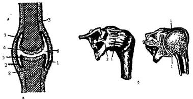
Rice. 2 Scheme of the structure of the joint:
1 - articular bag; 2 - inner shell of the joint;
3 - periosteum; 4 and 5 - articular cartilage; 6 - joint space;
7 - articular head; 8 - articular fossa;
On the upper limb There are the following main joints: shoulder joint; elbow joint; wrist joint.
On the lower limb the main joints are: hip joint, knee-joint and ankle joint.
The human skeleton is made up of four sections: the skeleton of the head (skull), the skeleton of the trunk, the skeleton of the upper and lower extremities.
Head skeleton. The bones of the head together make up the skull. With the exception of the lower part, the bones of the skull are firmly connected by sutures. They form receptacles for the brain and sense organs (vision, hearing, smell). The bones of the skull are the support for the initial sections of the respiratory tract (nasal cavity) and digestive system(skeleton of the oral cavity). The skull has in its structure: paired bones (temporal, parietal); unpaired (frontal, occipital); upper jaw and lower jaw. When examining the skull from the front, the cavities of the two eye sockets are visible, and between them is the entrance to the nasal cavity (piri-shaped opening).
The skeleton of the body includes the vertebral column and the bones that make up the chest. vertebral column – it is the support of the body, it withstands the weight of the head, trunk and upper limbs (2/3 of the body weight) and transfers it to the pelvis and lower limbs. In humans, the spinal column consists of 33 - 34 vertebrae. There are 5 sections of the spine: cervical, consisting of 7 vertebrae, thoracic - from 12, lumbar - from 5, sacral (sacrum) - from 5 and coccygeal (coccyx) - from 4 - 5 vertebrae. The vertebrae, with the exception of 1 - 2 cervical vertebrae, have a common structural plan. A vertebra consists of a body and an arch. The body and arch limit the vertebral foramen. The vertebral foramina of all vertebrae form the spinal canal, in which the spinal cord is located.
The sacrum consists of five vertebrae, closely connected to each other into a single whole. The junction of the sacrum with the fifth lumbar vertebra represents a protrusion facing forward - a cape. The human spine is characterized by the presence of curves. The bend, facing the bulge forward, is called lordosis. The bend, facing the bulge back - kyphosis. A person has 2 lordosis (cervical and lumbar) and 2 kyphosis (thoracic and sacral). People normally also have a slight bend of the spine to the side - scoliosis. It arises in connection with the greater development of the muscles of one of the halves of the body and its greater mass. The curves of the spine provide mitigation of shocks and tremors of the body when jumping, running, walking. In the spine, flexion and extension, tilts to the sides and rotation around the axis are possible.
Rib cage.Thoracic vertebrae, 12 pairs of ribs and an unpaired sternum (sternum) and their connections make up the skeleton of the chest. The sternum is a flat bone. It consists of three parts: the upper one - the handle, the middle one - the body, the lower one - the xiphoid process. Ribs represented by 12 pairs of narrow, long, curved flat bones. The rib has a head, neck and body. The first 7 ribs are connected to the sternum with cartilage. These are true edges, the next 5 pairs are called false. 8, 9, 10 pairs are connected to each other by their cartilages - underlying with overlying, they form a costal arch. The anterior ends of the 11th and 12th ribs lie freely in the soft tissues, they are called oscillating ribs.
In women, the chest is shorter and more rounded than in men. Due to the fact that the longer lower ribs are more curved than the short upper ribs, the movements of the chest during breathing occur unevenly. When inhaling, the upper sections of the chest expand up and to the sides (thoracic breathing), the lower sections - in the transverse (abdominal breathing).
Upper limb bones.The skeleton of the upper limbs is formed by the shoulder girdle and the skeleton of the free upper limbs. The skeleton of the shoulder girdle consists of 2 shoulder blades and 2 clavicles. The skeleton of the free upper limbs (arms) is formed by the humerus, two bones of the forearm (ulna and radius) and the bones of the hand (carpal bones, metacarpal bones, phalanges of the fingers). Each finger, with the exception of the thumb, consists of 3 phalanges. The thumb consists of only 2 phalanges. The main functions of the skeleton of the upper limb are the organ of grasping and feeling.
Bones of the lower limbs.The skeleton of the lower extremities includes the pelvic girdle and free lower extremities (legs).
Pelvic girdle bones: 2 ilium, 2 ischial and 2 pubic bones. The skeleton of the free lower limb is formed by the femur, patella, lower leg bones (tibia and fibula) and foot. The bones of the foot are divided into the bones of the tarsus, metatarsus, and bones of the toes (phalanges). Connecting with each other, the bones of the foot form an elastic arch, facing upwards with a bulge. Behind the foot rests on the calcaneal tubercle, and in front - on the heads of the metatarsal bones. The main functions of the bones of the skeleton of the lower limb are the organ of support and movement.
muscles - active part of the human locomotor apparatus. All skeletal muscles, as well as the muscles of the head, trunk and limbs, are composed of striated muscle tissue. The contractions of the striated muscles are subject to the will of the person, therefore such muscles are called voluntary muscles. . Due smooth muscle tissue muscular membranes of internal organs, blood and lymphatic vessels, and skin muscles are formed. The contraction of smooth muscles is not subject to the will of a person, therefore smooth muscles are called involuntary. The contractile part of the skeletal muscles, formed by muscle fibers, passes into the tendon at both ends. With the help of tendons, muscles are attached to the bones of the skeleton. The tendons are very strong and strong. When the muscles contract, they cause the bones to move: flexion, extension, adduction, abduction or rotation.
I am writing this work in order to get to know the human musculoskeletal system in more detail, to determine its function in the body and what it consists of.
Musculoskeletal system
The musculoskeletal system consists of bones of the skeleton with joints, ligaments and muscles with tendons, which, along with movements, provide the supporting function of the body. Bones and joints participate passively in movement, obeying the action of muscles, but play a leading role in the implementation of the supporting function. A certain shape and structure of the bones give them greater strength, the reserve of which for compression, expansion, bending significantly exceeds the loads possible during the daily work of the musculoskeletal system. For example, the human tibia can withstand a load of more than a ton during compression, and in terms of tensile strength it is almost as good as cast iron. Ligaments and cartilage also have a large margin of safety.
Skeleton
The skeleton is made up of interconnected bones. It provides our body with support and shape retention, and also protects the internal organs. An adult human skeleton consists of about 200 bones. Each bone has a certain shape, size and occupies a certain position in the skeleton. Part of the bones are interconnected by movable joints. They are driven by muscles attached to them.
Spine. The original structure that makes up the main support of the skeleton is the spine. If it consisted of a solid bone rod, then our movements would be constrained, devoid of flexibility and would deliver the same unpleasant sensations as riding a cart without springs on a cobblestone pavement.
The elasticity of hundreds of ligaments, cartilage layers and bends makes the spine a strong and flexible support. Thanks to this structure of the spine, a person can bend down, jump, somersault, run. Very strong intervertebral ligaments allow the most complex movements and at the same time create reliable protection for the spinal cord. It is not subjected to any mechanical stretching, pressure under the most incredible curves of the spine.
The bends of the spinal column correspond to the influence of the load on the axis of the skeleton. Therefore, the lower, more massive part becomes a support when moving; the upper, with free movement, helps to maintain balance. The vertebral column could be called the vertebral spring.
Wavy curves of the spine provide its elasticity. They appear with the development of the motor abilities of the child, when he begins to hold his head, stand, walk.
Rib cage. The thorax is formed by the thoracic vertebrae, twelve pairs of ribs, and a flat sternum, or sternum. The ribs are flat curved bones. Their rear ends are movably connected to the thoracic vertebrae, and the front ends of the ten upper ribs are connected to the sternum with the help of flexible cartilage. This ensures the mobility of the chest during breathing. The two lower pairs of ribs are shorter than the rest and terminate freely. The chest protects the heart and lungs, as well as the liver and stomach.
It is interesting to note that the ossification of the chest occurs later than other bones. By the age of twenty, the ossification of the ribs ends, and only by the age of thirty does the complete fusion of the parts of the sternum, consisting of the handle, the body of the sternum and the xiphoid process, occur.
The shape of the chest changes with age. In a newborn, it has, as a rule, the shape of a cone with the base turned down. Then the circumference of the chest in the first three years increases faster than the length of the body. Gradually, the cone-shaped chest acquires a rounded shape characteristic of a person. Its diameter is greater than its length.
The development of the chest depends on the lifestyle of a person. Compare an athlete, swimmer, athlete with a non-athlete. It is easy to understand that the development of the chest, its mobility depends on the development of the muscles. Therefore, in adolescents of twelve to fifteen years old who go in for sports, the circumference of the chest is seven to eight centimeters larger than that of their peers who do not go in for sports.
Improper seating of students at a desk, squeezing the chest can lead to its deformation, which disrupts the development of the heart, large vessels and lungs.
Limbs. Due to the fact that the limbs are attached to a reliable support, they have mobility in all directions and are able to withstand heavy physical exertion.
Light bones - clavicles and shoulder blades, lying on the upper part of the chest, cover it like a belt. This is the handhold. The protrusions and ridges on the collarbone and shoulder blade are the site of muscle attachment. The greater the strength of these muscles, the more developed the bone processes and irregularities. In an athlete, a loader, the longitudinal ridge of the scapula is more developed than in a watchmaker or accountant. The clavicle is the bridge between the bones of the trunk and arms. The shoulder blade and collarbone create a reliable spring support for the hand.
The position of the shoulder blades and collarbones can be used to judge the position of the hands. Anatomists helped to restore the broken off hands of the ancient Greek statue of Venus de Milo, determining their position by the silhouettes of the shoulder blades and collarbones.
The pelvic bones are thick, wide and almost completely fused. In humans, the pelvis justifies its name - it, like a bowl, supports the internal organs from below. This is one of the typical features of the human skeleton. The massiveness of the pelvis is proportional to the massiveness of the bones of the legs, which bear the main load when a person moves, therefore the human pelvic skeleton can withstand a large load.
Leg and hand. With a vertical posture, a person’s hands do not carry a constant load as supports, they acquire lightness and variety of action, freedom of movement. The hand can perform hundreds of thousands of different motor operations. The legs carry the entire weight of the body. They are massive, have extremely strong bones and ligaments.
The head of the shoulder has no restriction in wide circular movements of the arms, such as when throwing a javelin. The head of the femur protrudes deeply into the deepening of the pelvis, which limits movement. The ligaments of this joint are the strongest and hold the weight of the body on the hips.
Exercise and training achieve greater freedom of movement of the legs, despite their massiveness. A convincing example of this can be ballet art, gymnastics, martial arts.
The tubular bones of the arms and legs have a huge margin of safety. It is interesting that the location of the openwork crossbars of the Eiffel Tower corresponds to the structure of the spongy substance of the heads of tubular bones, as if J. Eiffel designed the bones. The engineer used the same laws of construction that determine the structure of the bone, giving it lightness and strength. This is the reason for the similarity of the metal structure and the living bone structure.
The elbow joint provides complex and diverse movements of the hand in the working life of a person. Only he has the ability to rotate the forearm around its axis, with a characteristic movement of unwinding or twisting.
The knee joint directs the lower leg when walking, running, jumping. The knee ligaments in humans determine the strength of the support when the limb is straightened.
The hand begins with a group of bones of the wrist. These bones do not experience strong pressure, perform a similar function, so they are small, monotonous, and difficult to distinguish. It is interesting to mention that the great anatomist Andrei Vesalius could, blindfolded, identify each carpal bone and tell whether it belongs to the left or right hand.
The bones of the metacarpus are moderately mobile, they are located in the form of a fan and serve as a support for the fingers. Phalanges of fingers - 14. All fingers have three bones, except for the thumb - it has two bones. A person has a very mobile thumb. It can become at right angles to everyone else. Its metacarpal bone is able to oppose the rest of the bones of the hand.
The development of the thumb is associated with the labor movements of the hand. The Indians call the thumb "mother", the Javanese - "big brother". In ancient times, captives were cut off the thumb to humiliate their human dignity and make them unfit for participation in battles.
The brush makes the most subtle movements. In any working position of the hand, the hand retains complete freedom of movement.
The foot became more massive due to walking. The tarsal bones are very large and strong compared to the carpal bones. The largest of them are the talus and calcaneus. They can withstand significant weight of the body. In newborns, the movements of the foot and thumb are similar to those of monkeys. Strengthening the supporting role of the foot during walking led to the formation of its arch. When walking, standing, you can easily feel how the entire space between these points “hangs in the air”.
The vault, as is known in mechanics, withstands greater pressure than the platform. The arch of the foot provides the elasticity of the gait, eliminates pressure on the nerves and blood vessels. Its formation in the history of the origin of man is associated with upright walking and is a distinctive feature of man, acquired in the process of his historical development.
Two types of muscle tissue.
Smooth muscles. When we talked about muscles, we usually thought of skeletal muscles. But, besides them, in our body in the connective tissue there are smooth muscles in the form of single cells, in some places they are collected in bundles.
Many smooth muscles in the skin, they are located at the base of the hair bag. By contracting, these muscles raise the hair and squeeze out fat from the sebaceous gland.
In the eye around the pupil are smooth circular and radial muscles. They work all the time, imperceptibly for us, work: in bright light, the circular muscles constrict the pupil, and in the dark, the radial muscles contract and the pupil expands.
In the walls of all tubular organs - the respiratory tract, blood vessels, digestive tract, urethra, etc. - there is a layer of smooth muscles. Under the influence of nerve impulses, it is reduced. For example, reducing it in the windpipe delays the flow of air containing harmful impurities - dust, gases.
Due to the contraction and relaxation of the smooth cells of the walls of blood vessels, their lumen either narrows or expands, which contributes to the distribution of blood in the body. The smooth muscles of the esophagus, contracting, push a lump of food or a sip of water into the stomach.
Complex plexuses of smooth muscle cells are formed in organs with a wide cavity - in the stomach, bladder, uterus. The contraction of these cells causes compression and narrowing of the lumen of the organ. The strength of each cell contraction is negligible, since they are very small. However, the addition of the forces of entire beams can create a contraction of enormous force. Powerful contractions create a sensation of intense pain.
Muscles of the skeleton. Skeletal muscles carry out both static activity, fixing the body in a certain position, and dynamic, ensuring the movement of the body in space and its individual parts relative to each other. Both types of muscular activity closely interact, complementing each other: static activity provides a natural background for dynamic activity. As a rule, the position of the joint is changed with the help of several muscles of multidirectional, including opposite action. Complex joint movements are performed by coordinated, simultaneous or sequential contraction of non-directional muscles. Consistency (coordination) is especially necessary for the performance of motor acts in which many joints participate (for example, skiing, swimming).
Skeletal muscles are not only the executive motor apparatus, but also a kind of sensory organs. In the muscle fiber and tendons there are nerve endings - receptors that send impulses to cells at various levels of the central nervous system. As a result, a closed cycle is created: impulses from various formations of the central nervous system, going along the motor nerves, cause muscle contraction, and impulses sent by muscle receptors inform the central nervous system about each element of the system. The cyclic system of connections ensures the accuracy of movements and their coordination. Although the movement of skeletal muscles is controlled by various sections of the central nervous system, the leading role in ensuring interaction and setting the goal of a motor reaction belongs to the cerebral cortex. In the cerebral cortex, the motor and sensory zones of the representations form a single system, with each muscle group corresponding to a certain section of these zones. Such a relationship allows you to perform movements, attributing them to environmental factors acting on the body. Schematically, the control of arbitrary movements can be represented as follows. The tasks and purpose of a motor action are formed by thinking, which determines the direction of attention and efforts of a person. Thinking and emotions accumulate and direct these efforts. The mechanisms of higher nervous activity form the interaction of psychophysiological mechanisms of movement control at various levels. Based on the interaction of the musculoskeletal system, deployment and correction of motor activity are provided. Analyzers play an important role in the implementation of the motor reaction. The motor analyzer provides the dynamics and interconnection of muscle contractions, participates in the spatial and temporal organization of the motor act. The balance analyzer, or vestibular analyzer, interacts with the motor analyzer when the position of the body in space changes. Vision and hearing, actively perceiving information from the environment, are involved in spatial orientation and correction of motor reactions.
The name "muscle" comes from the word "musculis", which means "mouse".
This is due to the fact that anatomists, observing the contraction of skeletal muscles, noticed that they seem to run under the skin, like mice.
A muscle is made up of muscle plexuses. The length of muscle plexuses in humans reaches 12 cm. Each such plexus forms a separate muscle fiber.
Numerous rod-shaped nuclei are located under the shell of the muscle fiber. Along the entire length of the cell stretches several hundred of the thinnest filaments of the cytoplasm - myofibrils, capable of contracting. In turn, myofibrils are formed by 2.5 thousand protein filaments.
In myofibrils, light and dark discs alternate, and under a microscope, the muscle fiber looks transversely striated. Compare the function of skeletal and smooth muscles. It turns out that striated muscles cannot elongate as much as smooth ones. But skeletal muscles contract faster than the muscles of the internal organs. It is therefore not difficult to explain why a snail or an earthworm, devoid of striated muscles, moves slowly. The swiftness of the movements of the bee, lizard, eagle, horse, and man is ensured by the speed of contraction of the striated muscles.
The thickness of the muscle fibers of different people is not the same. For those who go in for sports, muscle fibers develop well, their mass is large, which means that the contraction force is also large. The limited work of the muscles leads to a significant reduction in the thickness of the fibers and the mass of the muscles as a whole, and also entails a decrease in the force of contraction.
There are 656 skeletal muscles in the human body. Almost all muscles are paired. The position of the muscles, their shape, the method of attachment to the bones has been studied in detail by anatomy. The location and structure of the muscles is especially important for the surgeon to know. That is why the surgeon is first and foremost an anatomist, and anatomy and surgery are sisters. World merits in the development of these sciences belong to our domestic science, and above all to N.I. Pirogov.
Nerve connections in muscles. It is wrong to think that the muscle itself can contract. It would be difficult to imagine at least one coordinated movement if the muscles were uncontrollable. “Start up” the muscle in the course of nerve impulses. An average of 20 impulses per second enters one muscle. In each step, for example, up to 300 muscles take part, and many impulses coordinate their work.
The number of nerve endings in different muscles is not the same. There are relatively few of them in the thigh muscles, and the oculomotor muscles, which make subtle and precise movements all day long, are rich in motor nerve endings. The cortex of the hemisphere is unevenly connected with individual muscle groups. For example, large areas of the cortex are occupied by motor areas that control the muscles of the face, hand, lips, and foot, and relatively small areas are occupied by the muscles of the shoulder, thigh, and lower leg. The size of individual zones of the motor area of the cortex is proportional not to the mass of muscle tissue, but to the subtlety and complexity of the movements of the corresponding organs.
Each muscle has a double nerve subordination. One nerve sends impulses from the brain and spinal cord. They cause muscle contraction. Others, moving away from the nodes that lie on the sides of the spinal cord, regulate their nutrition.
The nerve signals that control muscle movement and nutrition are consistent with the nervous regulation of muscle blood supply. It turns out a single triple nervous control.
Muscles generate heat. Striated muscles are “engines” in which chemical energy is immediately converted into mechanical energy. The muscle uses 33% of the chemical energy for movement, which is released during the breakdown of animal starch - glycogen. 67% of the energy in the form of heat is transferred by the blood to other tissues and evenly warms the body. That is why in the cold a person tries to move more, as if warming himself up due to the energy that the muscles produce. Small involuntary muscle contractions cause tremors - the body increases the production of heat.
Strength and speed of muscle contraction. The strength of a muscle depends on the number of muscle fibers, on its cross-sectional area, the size of the surface of the bone to which it is attached, the angle of attachment and the frequency of nerve impulses. All these factors have been identified by special studies.
The strength of a person's muscles is determined by what load he can lift. Muscles outside the body develop strength several times greater than that which is manifested in human movements.
The working quality of a muscle is associated with its ability to suddenly change its elasticity. Muscle protein becomes very elastic during contraction. After contraction of the muscle, it again acquires its original state. Becoming elastic, the muscle holds the load, this manifests muscle strength. A human muscle for every square centimeter of section develops a force of up to 156.8 N.
One of the strongest muscles is the calf. It can lift a load of 130 kg. Every healthy person is able to "stand on tiptoe" on one leg and even lift an additional load. This load falls mainly on the calf muscle.
Being under the influence of constant nerve impulses, the muscles of our body are always tense, or, as they say, are in a state of tone - a long contraction. You can check the muscle tone for yourself: close your eyes with force, and you will feel the trembling of the contracted muscles in the eye area.
It is known that any muscle can contract with different strengths. For example, the same muscles are involved in lifting a small stone and a pound weight, but they expend different strength. The speed with which we can set our muscles in motion varies and depends on the training of the body. The violinist makes 10 movements per second, and the pianist - up to 40.
Fatigue and rest
Reasons for fatigue. Fatigue is an indicator that the body cannot work to its full potential. Why does muscle fatigue occur? For science, this question has long been unresolved. Various theories have been created.
Some scientists have suggested that the muscle is depleted from a lack of nutrients; Others said that her "suffocation" was coming, a lack of oxygen. It has been suggested that fatigue occurs due to poisoning, or clogging, of the muscle with toxic waste products. However, all these theories did not satisfactorily explain the causes of fatigue. As a result, there was an assumption that the cause of fatigue does not lie in the muscle. A hypothesis of nerve fatigue has been put forward. However, an outstanding Russian physiologist, one of the students of I. M. Sechenov, Professor N. E. Vvdensky proved by example that the nerve conductors are practically not fatiguable.
The path to unraveling the mystery of fatigue was opened by the Russian physiologist I. M. Sechenov. He developed the nervous theory of fatigue. He found that the right hand, after prolonged work, restored its working capacity, if during the period of its rest movements were made with the left hand. The nerve centers of the left hand, as it were, energized the tired nerve centers of the right hand. It turned out that fatigue is removed more quickly when the rest of the working hand is combined with the work of the other hand than with complete rest. With these experiments, I. M. Sechenov outlined ways to relieve fatigue and methods for their reasonable organization of rest, thereby realizing his noble desire to facilitate human work.
Statics and dynamics of the human body
Equilibrium conditions. Every body has mass and has a center of gravity. A plumb line passing through the center of gravity (line of gravity) always falls onto the support. The lower the center of gravity and the wider the support, the more stable the balance. So, when standing, the center of gravity is placed approximately at the level of the second sacral vertebra. The line of gravity is between both feet, inside the support area.
The stability of the body increases significantly if you spread your legs: the area of \u200b\u200bsupport increases. When the legs approach each other, the area of support decreases, and therefore stability also decreases. The stability of a person standing on one leg is even less.
Our body has great mobility, and the center of gravity is constantly shifting. For example, when carrying a bucket of water in one hand, for stability, you lean in the opposite direction, while extending the other arm almost horizontally. If you carry a heavy object on your back, the body leans forward. In all these cases, the line of gravity approaches the edge of the support, so the balance of the body is stable. If the projection of the center of gravity of the body goes beyond the area of support, the body will fall. Its stability is ensured by a shift in the center of gravity, corresponding to a change in the position of the body. To create a counterweight, the body leans in the direction opposite to the load. The line of gravity remains inside the area of support.
By performing various gymnastic exercises, you can determine how balance and stability are maintained if the center of gravity goes beyond the fulcrum.
Rope walkers, for greater stability, take a pole in their hands, which they tilt to one side or the other. Balancing they move the center of gravity to a limited support.
Sports are for everyone
Muscle training. Active physical activity is one of the prerequisites for the harmonious development of a person.
Constant exercises lengthen the muscles, develop their ability to stretch better. During training, muscle mass increases, muscles become stronger, nerve impulses cause muscle contraction of great strength.
Muscle strength and bone strength are interrelated. When playing sports, the bones become thicker, and accordingly developed muscles have sufficient support. The entire skeleton becomes stronger and more resistant to stress and injury. A good physical load is a necessary condition for the normal growth and development of the body. A sedentary lifestyle is harmful to health. Lack of movement is the cause of flabbiness and muscle weakness. Physical exercises, work, games develop working capacity, endurance, strength, dexterity and speed.
Labor and sport. Movements in work and sports are forms of muscular activity. Work and sport are interconnected and complement each other.
Two students came to the workshop, stood at the workbench for the first time. One is into sports, the other is not. It is easy to see how quickly an athlete learns labor skills.
Sport develops important motor qualities - agility, speed, strength, endurance.
These qualities are improved in work.
Labor and physical education help each other. They favor mental work. When moving, the brain receives an abundance of nerve signals from the muscles that maintain its normal state and develop. Overcoming fatigue during physical labor increases efficiency during mental activities.
Conclusion:
So, the musculoskeletal system plays an important role in human life. It consists of the bones of the skeleton with joints, ligaments and muscles with tendons, which, along with movements, provide the supporting function of the body. Physical exercise and sports increase the strength of bone tissue, contribute to a stronger attachment to the bones of muscle tendons, strengthen the spine and eliminate unwanted curvatures in it, contribute to the expansion of the chest and the development of good posture.
Exercises for physical culture have the purpose of preventive, corrective and tonic action.
The complexity of determining and combining specific physical exercises, the sequence of their implementation in the classroom make it necessary to take into account the complex nature of the impact of exercises on those involved.
Ministry of Education and Science of the Russian Federation
federal state budgetary
educational institution of higher professional education "Tambov University named after G.R. Derzhavin"
Department of Physical Education.
By discipline: Physical culture
Topic: Musculoskeletal system.
Completed by a 1st year student
Faculty of International Relations
Nesmeyanova Alina
Scientific adviser:
Department Assistant
physical education
Saikin Sergey Vitalievich
Introduction.
Main part
1. The purpose of writing the work.
2.Method of working on materials
3. The result of the work. Conclusion.
Bibliography.
List of used literature.
1. "Body reserves" B. P. Nikitin, L. A. Nikitina. 1990
2. "A book for reading on human anatomy, physiology and hygiene." I.D.
Zverev, 1983
3. "Russian power". Valentin Lavrov. 1991
4. "Secrets of athleticism." Yuri Shaposhnikov. 1991
5. "Biology Man Grade 9". A. S. Batuev. 1997