Musculoskeletal system The human body is a real miracle of nature. It supports all parts of the body in the right position, protects the vital organs and provides amazing mobility to the entire body. Belt upper limbs responsible for attaching the hands to
Clavicle and shoulder blade construction
The composition of the upper limb belt implies a construction of two shoulder blades, two clavicles and the skeleton of the limb itself. It is the belt of the upper limbs that creates the shape of a person's shoulders. The arms are connected to the body by the shoulder blades and collarbones, which form the girdle of the upper limbs. The shoulder blades are located at the top of the back, have a triangular shape, they are connected to the spine and ribs with the help of muscles. The shoulder blade is paired with the clavicle, and the clavicle is connected to the sternum and ribs. The clavicle has the appearance of a curved bone passing between the sternum and the outer angle of the scapula.
The skeleton of the upper limb girdle is built from the following parts:
- 2 collarbones;
- 2 blades;
- humerus;
- radial bones;
- ulna bones;
- wrists;
- metacarpal bones;
- phalanges of fingers.
Function of the upper limb belt
The main function that the belt of the upper limbs of a person performs is to create a strong and maneuverable support for the hands. Unlike the pelvic girdle, it is not rigidly connected to axial skeleton. The main bones of the girdle of the upper limbs: the clavicle, which forms a regular joint with the sternum, and the scapula is connected to the bones of the body with powerful muscles. As a result, the shoulders are actively involved in the movements of the hands, increasing the amplitude and, accordingly, the efficiency of work.
Upper belt bones x limbs of a person have a structure, like that of a vertebrate skeleton, and consist of 3 sections - the shoulder, forearm and hand. The muscles associated with this belt strengthen shoulder joint and are responsible for most of the movements of the hands. Shoulder blade - a wide plate in the form of a triangle, located behind chest from the back, is part of the belt of the upper limbs. It has a flat cavity of the shoulder joint, in which the head is placed humerus. The shoulder joint is relatively unstable, it provides the maximum range of motion, but it is sensitive to dislocations and other injuries.
Main bones of the hands
The humerus is presented in the form of a long girdle of the upper limbs, two rather long ulna and radius bones are attached to it. The humerus forms elbow joint with both bones, and the hand connects to only one of them - the wrist joint. The ulna is placed on the inside. All the bones of the hand are connected to each other thanks to the joints.
The main ones are:
- brachial;
- wrist;
- elbow.
The joints are very varied in movement, with an active mobility that led to the transformation of the forelimb, that is, the arm, in the course of evolution into an organ of labor. and the radius is more stable than the humerus, respectively, the movements are less free. Even stronger knuckles. The structure and the leg are very similar. Their main difference is the device of the hand, in which the thumb is located separately from the rest, which allows the hand to make grasping movements. Between wrist and metacarpal bone This finger is the only saddle joint in the human body, the movements in which are much freer than at the base of the first toe.
The structure of the elbow joint
The belt of the upper extremities includes the elbow joint, which consists of two parts: block-shaped and spherical. The first connects the protrusion of the humerus with the ulnar notch, it also provides flexion-extension movements with the hands. The spherical part connects the head of the humerus with the radial fossa. This allows for twisting of the forearm. In general, the joint is quite stable due to the large joint surface and strong ligaments. The radius is the main bone in the forearm. It forms a joint with the wrist. The ulna together with the radius forms the elbow joint.
The structure of the shoulder joint
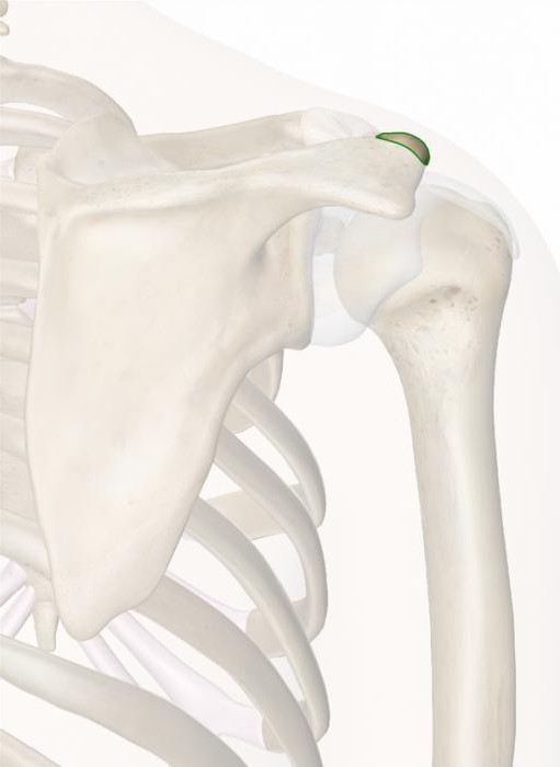
The girdle of the upper limbs includes the shoulder joint. The shoulder joint is the most mobile in the human body. Its almost flat cavity on the shoulder blade is articulated with the hemispherical head of the humerus. This device allows you to freely rotate your hand in all directions. The bone turns almost in a circle, up and down. Such mobility has its drawbacks, due to the fact that the strength of the connection is lowered, dislocations often occur in this joint. The second joint is formed by the scapula and collarbone. It often sprains when falling on an outstretched hand or when hit in front of the shoulder.
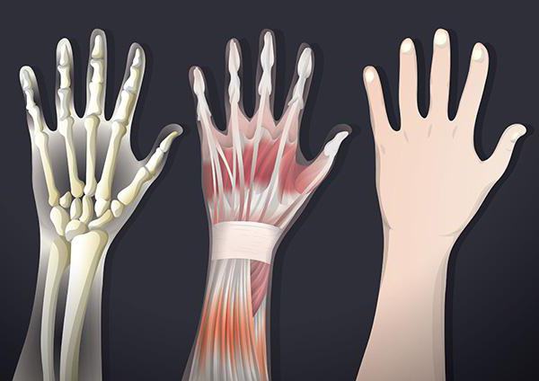
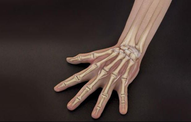
Wrist
This part of the hand has quite complex structure. The hand consists of 3 sections, which, in turn, have 27 bones. At the base of the palm are 5 metacarpal bones and 8 carpal bones. The skeleton of the fingers themselves is made up of 14 phalanges, 2 bones in the thumb and 3 in each of the four. has a highly specialized structure. In infants, they are only indicated, gradually forming, they will be clearly visible only by the age of seven, and their ossification is completed much later, by about 10-13 years. By the same period, the ossification of the phalanges of the fingers is completed.
Ligaments and muscles of the girdle of the upper limbs
Since the shoulder joint is quite mobile, and the shoulder girdle is not rigidly connected to the axial skeleton, the muscles of the girdle of the upper limb have a special function. Muscles connect the arm to the body and act as shock absorbers. The deltoid muscle is the largest and strongest in the shoulder, connecting the scapula and the humerus. It is thanks to her that the arm rises and moves back and forth.
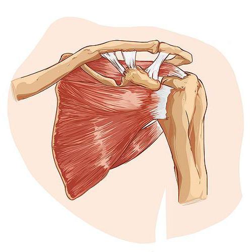
The rotator cuff is made up of four smaller muscles:
- infraspinatus;
- supraspinous;
- small round;
- subscapular.
They also control the rotation of the arm and strengthen the shoulder joint.
Major muscles of the girdle of the upper limb
The upper limb has a pair of main muscles: biceps and triceps, which form a pair of antagonists: if one contracts, the other of them relaxes. biceps, or biceps shoulder, goes from the scapula to the radius. If you bend your arm strongly, you can feel it well. Triceps, or connects the scapula to the ulna. It is not as noticeable, but larger than the biceps. When moving, they act as one muscle group. For example, when lifting the forearm, the biceps, the muscle that pulls the forearm toward the shoulder, contracts. At the same time, the triceps, an extensor muscle, is also stretched, which allows you to straighten your arm again.
Extensors and flexors
The complex movements of the wrist and hand are provided mainly by the coordinated work of many muscles passing through the forearm. These are flexors and extensors. The flexors bring the palm closer to the forearm and squeeze the fingers. They run along the inside of the arm. The extensors straighten the hand and fingers, bringing their back surface closer to the forearm. To open the palm and take an object with it, the coordinated work of 35 muscles of the forearm and hand is required. In addition, the muscles of the forearm deflect the hand to the left and right, rotate it, rotate the palm and fix the wrist and fingers in the desired position. Fine finger movements are controlled by the bone's own muscles, which run from the carpal bones to the base of the first phalanges. The work of the remaining phalanges is provided by long flexor and extensor tendons located in the forearm.
Age and prevention of bone aging
The human upper limb belt needs anti-aging prophylaxis. As we age, bone strength decreases and the risk of fractures increases. age loss bone tissue almost irreversible, but they can and should be prevented or significantly slowed down. Overstrain of the muscles of the shoulders and back is fraught with a very painful condition. People who spend all day at the computer and desk often slouch or hunch over. This results in stiffness of constantly tense muscles and stretching of the muscles of the shoulders and back, which threatens to cause painful muscle knots and tension headaches.
It is necessary to strengthen the upper limb belt, namely the muscles of the shoulders and back, through exercises that straighten the chest and unload the muscles and ligaments. It is very useful to reduce and dilute the shoulders, as well as shrugging the shoulders. Physical education relieves pain, strengthens muscles and bones, increases the flexibility of the body, and hence the mobility and work capacity of a person.
Skeleton top And bottom limbs has a general structural plan. Consists of two departments: skeleton belt And free limb skeleton.
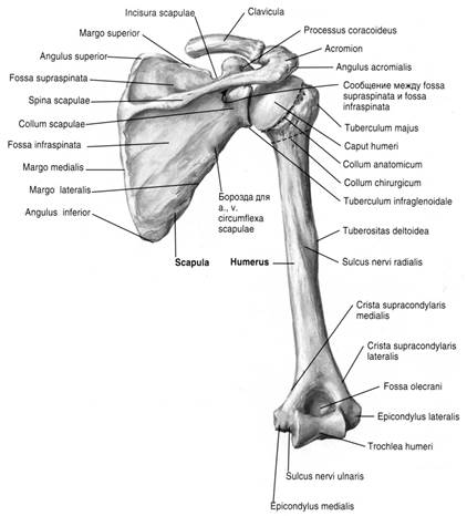
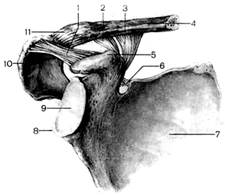
Bones shoulder girdle. The shoulder girdle consists of two bones: shoulder blades And clavicle .shoulder blade — flat triangular bone. Has three edges (superior, medial And lateral), three corners (medial, lateral And lower).
The shoulder blade is adjacent to the back of the chest. It is located at the level from II to VII ribs. The shoulder blade has an articular cavity for articulation with humerus and an anteriorly facing coracoid process for articulation with the clavicle. On the posterior surface of the scapula, a protrusion located transversely is visible.
Collarbone- this is an S-shaped curved tubular bone, having a body and two ends - sternal And acromial (brachial). The sternal end is thickened and connected to the handle of the sternum. The shoulder end is flat, articulates with the scapula.
Skeleton of the free upper limb comprises brachial bones, bones forearms (ulnar, radial bones) and brushes (bones wrists, metacarpuses and phalanges fingers).
Brachial bone — long tubular bone, has a body (diaphysis) and two ends (epiphysis). The upper end is represented by a rounded articular head for articulation with the scapula. It is separated from the body anatomic neck. Below the anatomical neck on the outside there are two elevations - big And small tubercles, separated by an intertubercular furrow. The narrowed part of the body closest to the head is called surgical neck. On the body of the humerus there is tuberosity, to which the deltoid muscle is attached. The lower epiphysis is expanded and ends condyle for articulation with the ulna and radius in the elbow joint
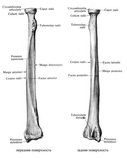
Forearm bones represented by two long tubular bones - the ulna and the radius
Elbow bone - located on the inside of the forearm from the side of the fifth finger (little finger). Top end ulna more massive, has two processes - the ulnar (behind) and the coronal (front), separated by a block-shaped notch for articulation with the humerus. The lateral (outer) surface of the coronoid process has a radial notch, which forms a joint with the articular circumference of the radius. The lower end of the ulna forms the head of the ulna. The head has an articular surface in the form of a circle for articulation with the ulnar notch of the radius. On the medial (inner) side is the medial styloid process.
Radius - a long tubular bone, located on the outer side of the forearm from the side of the I (thumb) finger. The upper end is formed by a cylindrical head, on which there is an articular fossa and an articular circumference. The upper ends of the ulna and radius are involved in the formation of the elbow joint. The lower end has a carpal articular surface, an ulnar notch, and a lateral styloid process. The lower surfaces of the ulna and radius are involved in the formation wrist joint with the upper row of carpal bones.
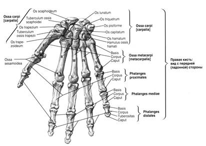
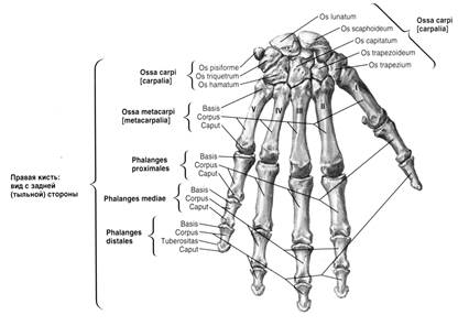
Hand bones represented by the bones of the wrist, metacarpal bones and bones (phalanges) of the fingers of the hand The wrist consists of eight short spongy bones arranged in two rows, 4 in each row. The bones of the wrist are articulated with each other. Top surface the upper row articulates with the carpal articular surface of the radius. Bottom row - with the bases of the metacarpal bones.
Metacarpal bones represented by 5 short tubular bones. They are counted from the side of the thumb (I, II, III, IV, V). Each metacarpal has a base, body and head, articulating with the upper phalanx of the corresponding finger.
Finger Skeleton formed by phalanges. Phalanges are short tubular bones in which the base, body and head are distinguished. The base and head have articular surfaces. The articular surface of the base at the upper phalanges articulates with the head of the corresponding metacarpal bone, at the middle and lower phalanges with the corresponding higher (proximal) phalanx.
Thumb has two phalanges. Each of the other fingers has 3 phalanges.
LECTURE №7.
1. Bones of the shoulder girdle, their connections.
2. Bones of the free upper limb and their connections.
3. Pelvic bones, their connections.
4. Bones free lower limb and their connections.
5. Typical sites of fractures of limb bones.
OBJECTIVE: To know the structure and connections of the bones of the upper and lower extremities.
To be able to show on bone preparations the components of the bones, bone protrusions, typical sites of fractures of the bones of the extremities. be able to distinguish female pelvis from the male.
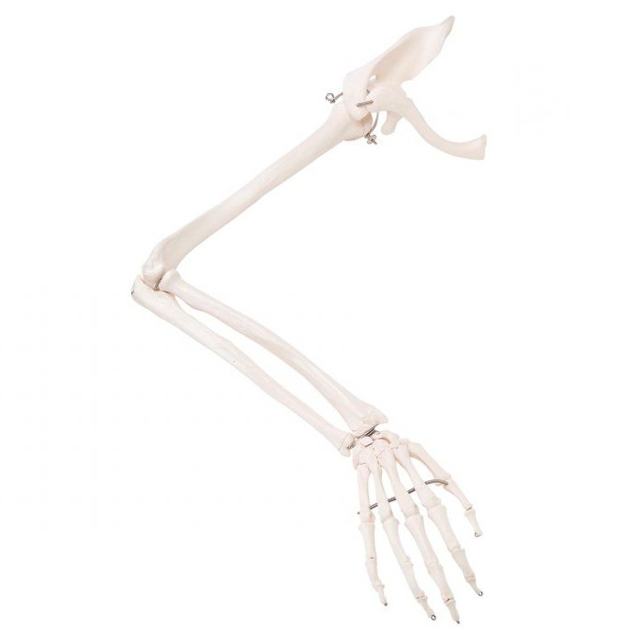
I. The functions of the human limbs are clearly delineated: the upper organs of labor, the lower ones - support and movement. But the upper and lower limbs have a common structural plan and consist of a belt and a free limb. The latter, in turn, is formed by three segments: the proximal one has one bone (humerus, femur), the middle one has two bones (radius, ulna and tibia, fibula) and the distal one has many bones (bones of the hand and foot).
The bones of the upper limb are divided into the girdle of the upper limb, consisting of the scapula and collarbone, and the skeleton of the free upper limb (hand), which includes the humerus, forearm [and ulna], carpal bones, metacarpal bones, and bones of the fingers (phalanges).
The clavicle (clavicula) is a steam room, S-shaped curved tubular bone, a body and two thickened articular ends are distinguished: the sternal and acromial. The medial part of the clavicle is convexly facing forward, and the lateral part is facing back.
Shoulder blade (scapula) - flat bone triangular shape. Three of its edges are distinguished: upper, lateral and medial and three corners: upper, lower and lateral. The lateral angle of the scapula is thickened and ends in an oval, shallow articular cavity for articulation with the humerus (spherical joint). With its anterior recessed surface, the scapula is adjacent to the posterior wall of the chest at the level of the II-VI ribs. On the back surface of the scapula there is a scapular spine, passing into the shoulder process - the acromion. The acromion has articular surface for articulation with the clavicle.
The sternal end of the clavicle articulates with the sternum, forming a saddle-shaped (or flat) sternoclavicular joint with an intra-articular disc that divides the joint cavity into two parts (chambers). The presence of the disk provides the possibility of movement in the joint around three axes. The sternoclavicular joint is the only joint that connects the girdle of the upper limb to the skeleton of the body. The lateral end of the clavicle is connected to the acromion of the scapula by a flat, inactive acromioclavicular joint.
2. Humerus (humerus) - a typical long tubular bone, has a body (diaphysis) and two ends (epiphysis). At the proximal end there is a head, separated from the rest of the bone by an anatomical neck. Below the anatomical neck, there is a small tubercle on the anterior surface, and a large tubercle on the lateral surface. Between them passes the intertubercular groove, intended for the tendon of the long head of the biceps brachii muscle. Below the large and small tubercles, at the point of transition of the upper epiphysis into the body, there is a slight narrowing - the surgical neck (fractures are more often observed in this place).
The distal end of the bone is thickened and is called the condyle of the humerus, consisting of the head of the condyle, with which the head of the radius articulates, and the block, which connects to the trochlear notch of the ulna at the elbow joint.
The radius (radius) is located on the outer surface of the forearm from the side of the thumb, the ulna (ulna) - on the inner surface from the side of the fifth finger (little finger). Both of these bones are tubular, trihedral. They consist of a body and two ends (epiphyses. The upper and lower epiphyses touch each other, forming the proximal and distal radioulnar joints, allowing the radius together with the hand to rotate (supination and pronation) by almost 180 °.
The bones of the hand (ossa manus) are divided into the bones of the wrist, metacarpal bones and bones of the fingers (phalanges).
The bones of the wrist (ossa carpi) are located in two rows of 4 each, counting from the thumb, the upper row includes the navicular, lunate, triquetral, pisiform bones; the lower row is made up of a bone - a trapezoid, a trapezoid, a capitate and an hamate. Three bones of the proximal row (with the exception of the pisiform) form an elliptical articular surface for articulation with the bones of the forearm. All bones of the wrist are spongy bones.
The metacarpal bones (ossa metacarpi) are represented by five short tubular bones, which are counted from the side of the thumb (I, II, III, etc.). Each metacarpal bone has a base, body, head. On the base and head there are articular surfaces for articulation with the bones of the wrist and phalanges of the fingers.
The bones of the fingers, or phalanges (ossa digitorum, seu phalanges), are formed by short tubular bones - phalanges: proximal, middle and distal (nail). The thumb consists of only two phalanges: proximal and distal.
Chapter 1. Topic: The skeleton of the upper limb.
Summary.
Classification of the bones that form the girdle of the upper limb and the free part of the upper limb. The structure of the bones of the shoulder girdle (collarbone, shoulder blade). The structure of the bones of the free part of the upper limb (humerus, bones of the forearm and hand). Basic morphological formations. Anatomical substantiation of the most common of their injuries.
Purpose and objectives of the lesson
Students should know:
1. The main morphological formations of the bones of the belt and the free upper limb.
2. Latin and Russian names.
3. Sites of frequent fractures on the ulna and radius (olecranon, distal ends).
Students should be able to:
1. Determine any individual bone of the skeleton of the upper limb.
2. Distinguish the bones of the right upper limb from the left.
3. Find parts of the bone - the body (diaphysis), ends (epiphysis), processes, etc.
4. Characterize the bone in shape - tubular (long or short, flat, spongy, mixed).
5. Feel the protruding bone points on yourself.
6. Freely navigate the diagrams and drawings from the textbook and atlas.
All bones should be studied on the skeleton, as well as using a set of individual bones, a textbook and atlas on human anatomy, and museum preparations (showcase No. 2).
In the skeleton of the human upper limb, the girdle of the upper limb and the free part of the upper limb are distinguished.
1. Upper limb belt (cingulum membri superioris) consists of two bones - the clavicle and the scapula.
shoulder blade (scapula)- a flat triangular bone. Adjacent to the back of the chest from the 2nd to the 7th rib.
In the shoulder blade distinguish:
edge medial (margo medialis), lateral (margo lateralis), superior (margo superior),
corners upper (angulus superior), lower (angulus inferior) and
lateral ( angulus lateralis)
costal surfaces (facies costalis), back (facies posterior )
On the back surface - awn ( spina scapule), which ends with the shoulder process ( acromion) and supraspinatus (fossa supraspinata) and infraspinatus (fossa infraspinata) fossa.
Rice. 1-1. the structure of the bones of the upper shoulder girdle (front view).
The coracoid process (processus coracoideus) departs from the upper edge
The lateral angle forms the articular cavity (cavitas glenoidalis) and: supraarticular (tuberculum cupraglenoidale) and subarticular (tuberculum infraglenoidale) tubercles.
Collarbone(clavicula ) s-shaped curved tubular bone. It is the only bone that connects the upper limb to the skeleton of the body, providing greater freedom of movement of the limb.
In the clavicle, there are: a body (corpus claviculae) and two ends: sternal (extremitas sternalis), acromial (extremitas acromialis)
The upper surface of the clavicle is smooth, while the lower surface is rough. Near the acromial end there are: a cone-shaped tubercle (tuberculum conoideum) and a trapezoid line (linea trapezoidea ) . Ligaments are attached to these tubercles.
Rice. 1-2. The structure of the bones of the upper shoulder girdle (rear view).
2. free part upper limb(pars libera membri superioris ) .
It is formed by: the humerus, bones of the forearm and hand.
Brachial bone(humerus ) belongs to the typical long tubular bones. Distinguish the body of the humerus (corpus humeri) and two ends - the upper (proximal) and lower (distal).
The body of the humerus in the upper part has the shape of a cylinder, becomes trihedral downwards.
At this level, the posterior surface (facies posterior) and two lateral surfaces are distinguished: the medial anterior (facies anterior medialis) and the lateral anterior (facies anterior lateralis). ).
Rice. 1-3. The structure of the bones of the forearm.
In the middle lateral part of the body of the bone is the deltoid tuberosity (tuberositas deltoidea), which is the site of attachment of the muscle of the same name. Below the deltoid tuberosity, on the posterior surface of the bone, there is a spiral groove of the radial nerve (sulcus nervi radialis).
The upper end forms the head of the humerus (caput humeri). A furrow runs along its edge - the anatomical neck (collum anatomicum).
Behind the neck are two tubercles (for attaching muscles). The large tubercle (tuberculum majus) lies laterally. The small tubercle (tuberculum minus) is located anterior to the large tubercle.
From each tubercle there are ridges for attaching muscles. Between the tubercles passes the intertubercular groove (sulcus intertubercularis), in which the tendon of the long head of the biceps brachii muscle is located.
The narrowest part of the bone, located between the head and the body, is called the surgical neck (collum chirurgicum), the site of the most frequent fractures.
The lower end is expanded and ends with the condyle of the humerus (condylus humeri). It distinguishes between a block (trochlea humeri), and a head (capitulum humeri) for articulation with the radius and ulna.
In front of the block is the coronary fossa (fossa coronoidea ) . Above the head of the condyle is the radial fossa (fossa radialis ) .
Behind - the fossa of the olecranon (fossa olecran i).
On the sides of the condyle are epicondyles: medial (epicondylus medialis) and lateral (epicondylus lateralis) for attaching muscles and ligaments. On the posterior surface of the medial epicondyle is the groove of the ulnar nerve (sulcus nervi ulnaris).
Forearm(antebracium ) consists of two bones: medially located ulna(ulna) and laterally radius(radius).
They are long tubular bones.
Each bone consists of a body and two ends. The bodies of both bones have a trihedral shape with three surfaces and edges.
One surface is turned back (facies posterior), the other - forward (facies anterior), the third - at the radius laterally (facies lateralis), at the ulna - medially (facies medialis).
Of the three edges, one is sharp and faces the adjacent bone, limiting the interosseous space. This is the interosseous edge ( margo interosseus). In addition to common features, each bone has characteristics buildings. At the proximal end of the ulna, there is a block-shaped notch (incisura trochlearis), an ulna (olecranon), and a coronoid (processus coronoideus) processes. At the distal end, the head of the ulna (caput ulnae), the styloid process (processus styloideus) from the medial side.
At the proximal end of the radius, there is a head (caput radii) with articular surfaces: a fossa (fovea articularis) and a circle (circumferentia articularis). At the distal end - the carpal articular surface (facies articularis carpalis) and the styloid process (processus styloideu s) from the lateral side.
Some bone formations are an important guide for the doctor, so you need to be able to feel on the shoulder blade - the spine, acromion, lower and medial angles.
On the clavicle - the body and ends. On the humerus - the head and epicondyles. Styloid process on the radius and ulna. Olecranon and head on the ulna.
Brush (manus) consists of the bones of the wrist (ossa carpi), metacarpal bones (ossa metacarpi) and bones of the fingers, phalanges (ossa digitorum, phalanges ) .
Rice. 1-4. The structure of the bones of the hand (front view).
. 
Rice. 1-4. The structure of the bones of the hand (back view).
Wrist consists of eight spongy bones arranged in two rows.
Proximal row (from the thumb): navicular ( os scaphoideum) lunate (os lunatum), trihedral (os triquetrum), pisiform (os pisiforme), which is a sesamoid bone, i.e. is located in the thickness of the tendon of the ulnar flexor of the wrist.
Distal row: trapezium (os trapezium), trapezoid (os trapezoideum), capitate (os capitatum), hook-shaped (os hamatum).
On the palmar surface, a carpal groove is formed, which is limited laterally by the tubercle of the navicular bone and the trapezium bone, medially by the hook of the hamate bone and the pisiform bone.
Metacarpal bones (ossa metacarpi (I-V), belong to short tubular bones. Each consists of a base, body and head.
Finger bones or phalanges (ossa digitorum, ph alanges ), are short tubular bones.
Each finger, except for the first (big) has three phalanges: proximal, middle and distal. The thumb has only two phalanges - proximal and distal.
Thumb, 1st (pollex, digitus primus).
Forefinger, 2nd (index, digitus secundus).
Middle finger, 3rd (digitus medius, tertius).
Ring finger, 4th (digitus anularis, quartus).
Little finger, 5th (digitus minimus, quintus)
Questions for self-control.
- On which part of the humerus is the deltoid tuberosity located?
- Where does the sulcus of the radial nerve pass?
- What is the condyle of the humerus formed by?
- What processes terminate the trochlear notch of the ulna?
- List on Latin finger names.
(Answer in writing in the workbook).
Choose one correct answer.
- Which of the following bones has a styloid process?
a) shoulder
b) elbow
c) clavicle
d) shoulder blade
e) sternum
- Which of the following bones belongs to the girdle of the upper limb?
a) shoulder blade
b) shoulder
c) radiation
d) elbow
- Which of the following bones are spongy in structure?
a) shoulder blade
b) clavicle
c) capitate bone
d) humerus
e) radius
- The proximal row of carpal bones includes the following bones, except
a) crescent
b) trihedral
c) pea
d) scaphoid
e) trapezoidal
- The distal carpal bones include the following bones except
a) trapezoid
b) trapezoidal
c) capitate
d) hooked
e) pea-shaped
- The carpal groove is bounded by the following bones except
a) hook of the hamate
b) tubercle of the navicular bone
c) tubercle of the trapezoid bone
d) pea-shaped
e) capitate
- The first metacarpal has an articular surface at its proximal end for articulation with
a) triangular
b) capitate
c) trapezoidal
d) bone - trapezium
e) scaphoid
- On which formation of the humerus is the groove of the ulnar nerve located?
a) on the medial epicondyle
b) on the lateral epicondyle
c) on the block of the humerus
d) on the head of the condyle of the humerus
e) on the greater tubercle of the humerus
Equipment.
Skeleton. Separate bones of the skeleton of the upper limb. Museum natural anatomical preparations (showcase No. 2).
Literature.
- Human anatomy. // Ed. M.R. Sapina - M.: Medicine, Vol. 1, 1993, pp. 123-126, 126-133.
- Human anatomy. // Ed. M.G. weight gain. - M.: Medicine, 1999
- Sinelnikov R.D., Sinelnikov Ya.R. Atlas of human anatomy.-M.: Medgiz, T.1, 1989. p.82-84, p.84-100.
Goals and objectives of the lesson.
Students should know:
1. Types of continuous joints of the bones of the skull (syndesmosis, synchondrosis, synostosis).
2. The structure of the temporal mandibular joint, motion features.
3. Features of movement in the temporomandibular joint
Students should be able to:
1. Navigate in natural anatomical preparations, show the main morphological formations.
2. Be able to distinguish different types joints of the bones of the skull.
The bones of the skull are interconnected by means of continuous joints and discontinuous joints (temporomandibular joint).
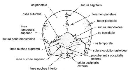
Rice. 2-1. Skull (cranium) rear view.
Continuous connections of the bones of the skull are represented by:
1. Fibrous connections in the form of seams,
2. Cartilaginous joints - synchondrosis (on the base of the skull), which turn into synostoses with age.
Seams are - jagged, scaly and flat.
1. The bones of the roof of the skull are connected to each other using serrated and scaly sutures.
The medial edges of the parietal bones are connected by a serrated suture called sagittal (sutura sagittalis).
The frontal bone is connected to the parietal by a jagged suture called coronal suture (sutura coronalis).
The parietal and occipital bones are joined by a serrated suture called lambdoid suture (sutura lambdoidea).
Scales temporal bone connects with parietal bone and greater wing sphenoid bone with a scalloped seam.
Bones facial skull connected with flat seams. The names of individual sutures on the skull are derived from the names of the connecting bones, for example sutura temporozygomatica.
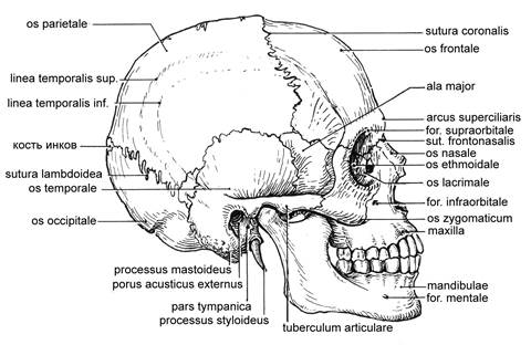
Rice. 2-2. Skull side view.
2. Synchondrosis - connection of bones with the help of cartilage (fibrous). There are wedge-occipital synchondrosis and petrooccipital synchondrosis. In place of the wedge-occipital synchondrosis, synostosis is formed with age.
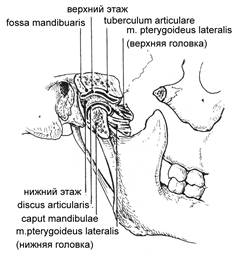
Temporomandibular joint (art. temporomandibularis) The joint is paired, complex elliptical.
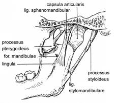
Rice. 2-3. Temporomandibular joint articulatio temporo-mandibularis, inside view.
Rice. 2-4. Temporomandibular joint articulatio temporo-mandibularis external view.
Attachment of the joint capsule. On the temporal bone, it is attached anterior to the articular tubercle, posteriorly at the level of the petrotympanic fissure. On the condylar process, the capsule is attached in front along the edge of the head, behind 0.5 cm below the posterior edge of the head mandible. The articular cavity is divided by cartilage into two isolated stages: upper and lower. Accordingly, the superior synovial membrane and the inferior synovial membrane are isolated.
Ligaments of the joint. On the lateral side - lateral ligament (lig. laterale). Starts from the base zygomatic process temporal bone, is attached to the posterolateral surface of the neck of the condylar process.
Sphenomandibular (lig. sphenomandibuiare) is located on the medial side of the joint. It starts from the spine of the sphenoid bone and is attached to the uvula of the lower jaw.
Awl-mandibular ligament (lig. stylomandibulare). It starts from styloid process, attached to the inner surface of the angle of the lower jaw.
Since the movements in the joints (right and left) pass together, they are functionally combined into a combined joint.
Functions:
1. Raising and lowering the lower jaw; that is, movement along the frontal axis.
2. movement of the lower jaw to the right and left
3. forward displacement of the lower jaw
When lowering the lower jaw, three phases are distinguished:
1. slight lowering of the lower jaw. The movement occurs in the lower floor of the joint, the disk remains in the articular fossa
2. significant lowering of the lower jaw. The cartilaginous disk, together with the head of the lower jaw, slides forward and exits onto the articular tubercle.
3. maximum lowering of the lower jaw. Movement occurs only in the lower floor around the frontal axis. The articular disc is located on the articular tubercle.
When the lower jaw is moved forward, movements occur only in the upper floor of the joint. When the lower jaw moves to the right in the left joint, the head slides with the disc in the upper floor. At this time, in the right temporomandibular joint, the head rotates around a vertical axis. When the lower jaw is displaced to the left, the head slides in the right joint, vertical rotation in the left.
Questions for self-control
1. Name the fibrous connections between the bones of the skull
2. List synchondrosis at the base of the skull
3. Phases in the movement of the temporomandibular joint
4. Name the ligaments of the temporomandibular joint
Test tasks
Exercise: choose one correct answer.
1. The parietal bones are connected using
A) coronal suture.
B) sagittal suture.
B) synchondrosis.
D) synostosis.
D) lambdoid suture.
2. The frontal bones are connected to the parietal with the help of
A) sagittal suture.
B) lambdoid suture.
B) coronal suture.
D) fronto-parietal suture.
D) synchondrosis.
3. The occipital bone is connected to the parietal with:
A) occipital-parietal suture.
B) parieto-occipital suture.
B) lambdoid suture.
D) coronal suture.
D) sagittal suture.
4. The ligaments of the temporal mandibular joint include
A) lateral ligament.
B) medial ligament.
B) collateral ligament.
D) yellow ligament.
D) longitudinal ligament.
5. The ligaments of the temporomandibular joint include:
A) medial ligament.
B) collateral ligament.
B) wedge-parietal ligament.
D) sphenomandibular ligament.
D) wedge-maxillary ligament.
Choose one wrong answer:
6. The joints of the bones of the skull include all of the following, except:
A) sagittal suture.
B) coronal suture.
B) temporomandibular joint.
D) petrooccipital synchondrosis.
D) atlanto-occipital connection.
7. The temporomandibular joint belongs to all of the listed formations, except:
A) articular disc.
B) lateral ligament.
B) collateral ligament.
D) head of the lower jaw.
D) mandibular fossa.
8. For the temporomandibular joint, all statements are true, except:
A) complex joint.
B) combined joint.
B) ellipsoid joint.
D) biaxial joint.
D) triaxial joint.
9. For the temporomandibular joint, all statements are true except:
A) combined joint.
B) uniaxial joint.
B) biaxial joint.
D) movements take place in the frontal and vertical axis.
E) movements take place in the frontal and sagittal axis.
10. The joints of the bones of the skull include all of the following, except:
A) lambdoid suture.
B) coronal suture.
B) injections.
D) synchondrosis.
D) synostosis.
Equipment
Museum stand No. 14, wet preparation "temporomandibular joint", skull.
Literature
1. "Human Anatomy" Volume 1, edited by M.R. Sapina. Publishing house "Medicine" 1996. pp.162-165.
2. "Atlas of human anatomy" volume 1, edited by R.D. Sinelnikov. Publishing house "Medicine" 1989. Page 149-151, pp. 35.
3. "Atlas of human anatomy" edited by F. Netter. Publishing house GEOTAR-Med 2003 Fig. 2-3.11.
Goals and objectives of the lesson
Students should know:
1. The structure of the sternum - clavicular joint, acromioclavicular joint of the shoulder joint.
2. Features of movement in the listed joints.
Students should know:
1. The main formations of these joints, as well as periarticular formations.
2. Features of movement in the sternoclavicular joint, acromioclavicular joint.
3. Features of movement in the shoulder joint, features of fixation of the shoulder joint and the clinical significance of this fixation.
The joints of the girdle of the upper limb (articulationes cinguli membri superioris):
1. Sternoclavicular joint
2. Acromioclavicular joint
sternoclavicular joint (articulatio sternoclavicularis). The joint is formed by the sternal articular surface of the clavicle and the clavicular notch of the sternum. The joint is complex, because inside the joint there is an articular disc that divides the articular surface into two chambers. The shape approaches the saddle or flat. It is a multiaxial joint.
Ligaments that strengthen the joint:
1. Anterior sternoclavicular ligament (lig. sternoclaviculare anterius).
2. Posterior sternoclavicular ligament (lig. sternoclaviculare posterius), which is a thickening of the fibrous membrane of the joint capsule in front and behind.
3. Interclavicular ligament (lig. Intraclaviculare), located above the jugular notch of the sternum.
4. The costoclavicular ligament (lig costoclaviculare) is located at some distance from the joint.
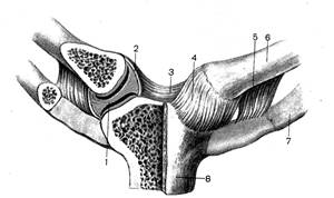
Rice. 3-1. Sternoclavicular joints, artt. sternoclaviculares; front view. (1.- discus articularis; 2.- capsula artcularis; 3.- lig. interclaviculare; 4.- lig. sternoclaviculare anterius; 5.- lig. costoclaviculare; 6.- clavicula; 7.- costa 1; 8.- manubrium sterni.
Acromioclavicular joint (articulatio acromioclavicularis). Formed by the acromion of the scapula and the acromial end of the clavicle. Inside the joint there is cartilage (discus articularis). The joint is flat, inactive, multiaxial.
Rice. 3-2. Acromioclavicular joint art. acromyoclavicularis; right. (1.- lig. coracoacromiale; 2.- lig. trapezoideum; 3.- lig. conoideum; 4.- extremitas acromialis claviculae; 5.- processus coracoideus; 6.- lig. transversum scapulae superius; 7.- scapula; 8 .- labrum glenoidale; 9.- cavitas glenoidalis; 10.- acromion; 11.- art. acromioclavicularis;
Ligaments that strengthen the joint:
1. Acromioclavicular ligament (lig. acromioclaviculare) - thickening of the joint capsule.
2. Coracoclavicular ligament (lig. coracoclaviculare) - away from the joint and consists of two bundles:
a) trapezoid ligament (lig. trapezoideum) - located laterally
b) conical ligament (lig.conoideum) - located medially.
Own ligaments of the scapula:
1. The coracoacromial ligament (lig. coracoacromiale) extends in the form of a vault over the shoulder joint from the anterior edge of the acromial process to the processus coracoideus.
2. The superior transverse ligament of the scapula (lig. transversum scapulae superius) is stretched over the scapular notch and turns it into a hole.
3. The lower transverse ligament of the scapula (lig. transversum scapulae inferius) runs from the base of the acromion through the neck of the scapula to the posterior edge of the articular cavity; passes under it. suprascapularis.
Questions for self-control
1. Features of the structure of the shoulder joint
2. The structure of the sternoclavicular joint
3. The structure of the acromioclavicular joint
Test tasks
Choose one wrong answer.
1. All formations belong to the sternoclavicular joint, except:
A) the articular surface of the sternal end of the clavicle.
B) clavicular notch of the manubrium of the sternum.
B) radiant ligament.
D) interclavicular ligament.
D) sternoclavicular ligaments.
2. All of the following structures belong to the shoulder joint, except:
A) heads of the humerus
B) articular cavity of the scapula
B) articular lip
D) coracobrachial ligament
D) acromioclavicular ligament
3. The acromioclavicular joint is strengthened by all of the listed ligaments, except:
A) acromioclavicular ligament
B) coracoclavicular ligament
B) trapezius ligament
D) conical ligament
D) deltoid ligament
4. Own ligaments of the scapula include all, except:
A) superior transverse ligament
B) middle transverse ligament
B) lower transverse ligament
D) coracoid-acromial ligament
5. For the shoulder joint, all statements are true except:
A) ball joint
B) multiaxial joint
C) the articular surfaces of the scapula and shoulder are complemented by the articular lip
D) has a coracobrachial ligament
D) has an acromio-brachial ligament
Equipment
Museum showcase No. 14, wet preparations "sternoclavicular joint", "acromio-clavicular joint", "shoulder joint"
Literature
4. "Human Anatomy" Volume 1, edited by M.R. Sapina. Publishing house "Medicine" 1996. pp.180-186
5. "Atlas of human anatomy" volume 1, edited by R.D. Sinelnikov. Publishing house "Medicine" 1989. Page 151-154
6. "Atlas of human anatomy" edited by F. Netter. Publishing house GEOTAR-Med 2003 Fig.394.
Purpose and objectives of the lesson.
To study the muscles of the head, their origin, place of attachment and function. Examine the fasciae of the head.
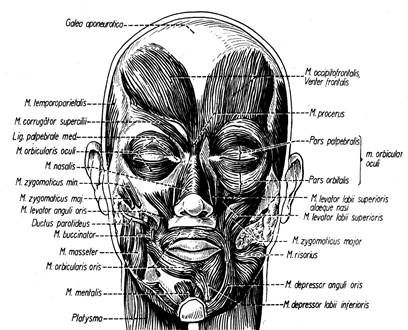
The muscles of the head and face are divided into mimic and chewing.
Mimic muscles:
They develop from the mesenchyme of the second visceral arch.
They don't have fascia.
They have at least one point of attachment to the skin.
Innervated by the facial nerve.
Reflect the emotional state of a person.
Among the mimic muscles, the mimic muscles of the head and face are distinguished
Mimic muscles of the head (cranial vault).
1. M. occipitofrontalis, occipital-frontal muscle,
beginning: linea nuchae superior;
attachment: eyebrow skin;
has two abdomens: occipital (venter occipitalis) and frontal (venter frontalis), connected by a tendon aponeurosis (helmet - galea aponeurotica)
function: when contracting, horizontal folds appear on the skin of the forehead, the upper eyelid and eyebrow rise. The face acquires an expression of attention and mental tension.
2.M. temporoparietalis, temporoparietal muscle,
origin: inner surface of the cartilage of the ear;
attachment: fan-shaped to the lateral surface of galea aponeurotica.
Function: gives the face an expression of interest: "ears on top".
3. M. Procerus, proud muscle
Origin: bony dorsum of the nose;
Attachment: skin of a glabella;
Function: when it contracts, a skin fold appears, passing transversely to the root of the nose
Rice. 4-2. Muscles of the head
Mimic muscles of the face.
According to the location and function, the mimic muscles of the face are divided into:
muscles surrounding the palpebral fissure;
muscles surrounding the oral fissure;
muscles surrounding the nostrils and
ear muscles.
The circular muscles act as sphincters, the radial muscles act as dilators.
Mimic muscles of the orbital region.
1. M. orbicularis oculi, circular muscle of the eye:
Surrounds the palpebral fissure, located in the peripheral part of the pars orbitalis, on the bony edge of the orbit, and the inner part, pars palpebralis, on the eyelids. The smaller pars lacrimalis arises from the wall of the lacrimal sac, and by expanding it, affects the outflow of tears. Pars palpebralis closes the eyelids, and pars orbitalis closes the eye.
2.M. corrugator supercilii, muscle wrinkling the eyebrow:
Beginning: medial part of the superciliary arch;
Attachment: eyebrow skin;
Function: pulls the skin of the forehead down and medially, forming vertical folds above the root of the nose.
Muscles of the nose.
1. M. nasalis, nasal muscle
Consists of two parts: pars transversa, and pars alaris. Pars transversa, by narrowing the openings of the nostrils, directs air to the olfactory region of the nasal cavity (“sniffing”). It starts from the maxillae in the fossa canina area, goes perpendicular to the back of the nose, connects to the muscle of the other side. Pars alaris starts from the maxillae below and medially and is woven into the skin of the alar of the nose, dilates the nostrils, allowing deep breathing.
2. M. depressor septi nasi, the muscle that lowers the nasal septum:
Origin: above the medial incisor maxillae;
Insertion: cartilaginous part of the nasal septum;
Function: pulls down the nasal septum.
Muscles of the auricle
1. M. auricularis anterior, anterior ear muscle
Origin: temporal fascia and tendinous helmet anterior to the ear;
Attachment: ear skin;
Function: pull auricle forward.
2. M. auricularis superior, superior ear muscle:
Beginning: tendinous helmet up from the ear
Attachment: top part cartilage of the auricle;
Function: pulls the auricle up.
3. M. auricularis posterior, inferior ear muscle
Origin: processus mastoideus
Attachment: back surface of an auricle;
Function: pulls the auricle back.
Mimic muscles of the mouth
1. M. orbicularis oris, the circular muscle of the mouth forms the muscular basis of the upper and lower lips; consists of two parts pars marginalis (marginal) and pars labialis (labial). Pars labialis lies in the thickness of the lips, attaches to the corners of the mouth, where it is woven into the skin and mucous membranes. Pars marginalis is located at the corners of the mouth, where other muscles surrounding the oral fissure are woven into it;
Function: closes the oral fissure, participates in sucking, chewing, articulation.
2. M. depressor anguli oris, the muscle that lowers the corner of the mouth:
Beginning: lower edge of mandibulae;
Attachment: corner of a mouth;
Function: pulls the corner of the mouth down, gives the face an expression of sorrow.
3. M. depressor labii inferioris, the muscle that lowers the lower lip:
Beginning: the outer surface of the body of the lower jaw;
Insertion: skin of the lower lip;
Function: pulls the lower lip down and sideways
4. M. Mentalis, chin muscle
Origin: jugum alveolare of the medial incisor;
Attachment: skin of a chin;
Function: lifts the skin of the chin, protruding the lower lip.
5. M. Buccinator, buccal muscle:
Beginning: crista buccinatoria mandibulae, posterior edge of the alveolar process of maxillae and on a special sheer tendon cord, raphe buccopharyngea, located between maxillae and mandibulae.
Attachment: woven into the circular muscle of the mouth;
Function: forms the muscular base of the cheek, pulls the corner of the mouth back, presses the cheek to the teeth. Trumpeter's Muscle.
6. M. levator labii superioris, levator muscle upper lip
Beginning: infraorbital region upper jaw;
Attachment: upper lip;
Function: lifts the upper lip, forms the nasolabial furrow..
7. M. zygomaticus minor, small zygomatic muscle
Start: zygomatic bone
Attachment: nasolabial fold;
Function: Raises the corner of the mouth.
8. M. Zygomaticus major, large zygomatic muscle:
Origin: zygomatic bone;
Attachment: corner of a mouth;
Function: pulls the corner of the mouth up and out, a typical laughing muscle.
9. M. levator anguli oris, the muscle that raises the corner of the mouth:
Beginning: in the depths of fossa canina;
Attachment: skin of a corner of a mouth;
Function: Raises the corner of the mouth.
10. M. risorius, laughter muscle:
Beginning: chewing fascia;
Attachment: skin of a corner of a mouth;
Function: smile muscle, forms a dimple on the cheek.
M. Platysma (superficial muscle of the neck) should also be referred to mimic muscles in terms of origin, function, structural features, and innervation.
Origin: superficial plate of the thoracic fascia below the clavicle;
Attachment: the edge of the lower jaw and the corner of the mouth;
Function: lifts the skin of the neck, pulls the corner of the mouth down.
Chewing muscles.
Develop from the mesenchyme of the first visceral arch (mandibular)
They originate on the bones of the skull and attach to the lower jaw.
Innervated by the trigeminal nerve.
1. M. masseter, chewing muscle
beginning: lower edge of the zygomatic bone and zygomatic arch;
attachment: outer side of the branch of the lower jaw;
2. M. temporalis, temporalis muscle
beginning: temporal fossa of the skull to linea temporalis;
attachment: processus coronoideus mandibulae;
function: presses the lower teeth to the upper ones, horizontal beams push the lower jaw back.
3M. pterygoideus medialis, medial pterygoid muscle
origin: fossa pterygoidea of the pterygoid process of the sphenoid bone;
attachment: tuberositas pterygoidea mandibulae;
function: presses the lower teeth to the upper.
4. M. pterygoideus lateralis, lateral pterygoid muscle
start: bottom surface big wing and the pterygoid process of the sphenoid bone;
attachment: collum mandibulae, bag and articular disc of the temporomandibular joint;
function: with unilateral contraction, it shifts the lower jaw to the side; with bilateral contraction, it pushes the lower jaw forward; pulls the joint capsule and articular disc forward.
Fascia of the head
There are no fasciae in the facial area, since the facial muscles lie directly under the skin. The exception is M. Buccinator, covered in the back with a dense fascia buccopharyngea, which loosens anteriorly, merging with the tissue of the cheek, and fuses with the raphe buccopharyngea behind and continues into the connective tissue cover of the pharynx.
Fascia temporalis covers the muscle of the same name, starts at the top of the linea temporalis, and below is attached to the zygomatic arch with two plates to the outer and inner sides arcs.
Fascia massaterica covers the chewing muscle, is attached above to the zygomatic arch, and below to the edge of the lower jaw. Posteriorly goes into fascia parotidea covering the parotid salivary gland.
Questions for self-control.
1. In which direction does the lower jaw move during contraction of the right lateral pterygoid muscle?
2. which one chewing muscles drops the lower jaw?
3. List the features of M. Platysma that are common to other mimic muscles.
Test tasks.
1. draw 5 schematic facial expressions (like emoticons) and describe the tension of which facial muscles gives the face such an expression.
2. Present a description of the masticatory muscles (name, origin, attachment, function, fascia) in the form of a table.
3. in the form of a table, present the movements of the lower jaw and the muscles that carry them out.
1. Human anatomy, ed. M.R. Sapina, Moscow, Medicine, 1993, T.1., S.283-295.
2. Human anatomy. // Under. ed. M.G. Weight gain.-M.: Medicine, 1999, p.
3. Sinelnikov R.D., Sinelnikov Ya.R. Atlas of human anatomy.- M.: Medgiz, V.1, 1990. P.
Muscles acting on the joints of the shoulder girdle and the free upper limb.
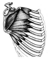
- M. pectoralis major, pectoralis major:
Beginning: from the clavicle (pars clavicularis), sternum and cartilage of the II-VII ribs (pars sternocostalis), sheath of the rectus abdominis muscle (pars abdominalis);
Attachment: crista tuberculi majoris humeri;
Function: brings the hand to the body and turns it inward; with fixed arms, it acts as an inspiratory muscle.
 | Rice. 5-1. M. pectoralis major |
- M. pectoralis minor pectoralis minor muscle
Beginning: II-V ribs;
Insertion: coracoid process of the scapula;
Function: tilts the scapula forward and down, with a strengthened scapula, raises the ribs.
- M. subclavius, subclavian muscle
Beginning: cartilage of the 1st rib;
Insertion: acromial end of clavicle;
Function: strengthens the sternoclavicular joint.
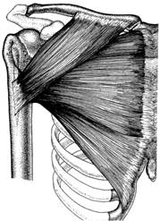
- M. Serratus anterior, the anterior serratus muscle is adjacent to the side of the chest, forms the medial wall of the axillary cavity.
Beginning: teeth from nine top edges;
Attachment: medial edge of the scapula
Function: pulls the scapula laterally and upward, turning it upward with the glenoid cavity, which allows you to raise your hand above the horizontal.
 | Rice. 5-2. 4. M. Serratus anterior, |
Autochthonous (own) chest muscles
- mm. intercostales externi, the external intercostal muscles perform the intercostal spaces behind from the tubercle of the rib to the cartilage of the rib in front, where they continue into membrana intercostalis externa. They consist of flat short muscle bundles mixed with tendon bundles. Their fibers go from top to bottom and forward.
Beginning: the lower edges of the ribs from the tubercles of the ribs to the beginning of the cartilaginous part;
Attachment: the upper edge of the underlying rib.
Function: raise the ribs.
- mm. intercostales interni, the internal intercostal muscles also fill the intercostal spaces from the sternum in front to the angle of the ribs behind, where they continue into membrana intercostalis interna. They lie medially from the external muscles so that the groove on the lower edges of the ribs is between the external and internal intercostal muscles. Fibers mm. intercostals interni are directed from top to bottom and backwards, i.e. perpendicular to the fibers of the external intercostal muscles.
Beginning: upper edge of the rib;
Attachment: the lower edge of the overlying rib.
Function: lower ribs.
SKELETON LIMB
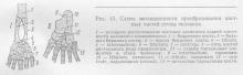
locomotion (locomotion) most vertebrates are primarily associated with limbs, which reach full development in terrestrial forms that raise the body above the ground. In this case, the limbs take a vertical position.
The prototype of the limbs of vertebrates are the paired fins of fish, which consist of cartilaginous rays and represent a simple flexible lever formed under the influence of movement in a liquid medium. In terrestrial, in connection with the conditions of existence, the fin is transformed into a five-fingered limb.
The skeleton of the limbs consists of two sections: the skeleton of the free limb and the so-called belts, shoulder and pelvic, through which the limbs are attached to the body. From the dorsal part of the primitive shoulder girdle, a scapula is formed, bearing a hole for articulation with the skeleton of the free upper limb; a coracoid arises from the ventral part, which in amphibians, reptiles and birds adjoins the sternum. Cranial to the coracoid is another process, the procoracoid, which is replaced by the integumentary bone, the clavicle, that develops in its place. This latter, connecting with the sternum, enters into communication with the scapula. In viviparous mammals, the coracoid is reduced, loses its connection with the sternum and adheres to the scapula in the form of its coracoid process, processus coracoideus. The scapula in these animals is equipped with a ridge that continues into the acromial process, to which the clavicle is attached. The clavicles are developed in those forms of mammals whose limbs can move in all directions (many rodents, bats, monkeys and humans). In forms with limbs that perform monotonous movements in one plane when running, swimming, etc. (ungulates, carnivores, cetaceans), the clavicles are completely reduced.
The pelvic girdle in its primitive form in lower fish is represented by a plate lying on the ventral side of the body, to which both hind fins are attached. The dorsal part of the lateral half of the pelvic girdle, corresponding to the scapula of the shoulder girdle, in terrestrial vertebrates forms ilium, ilium; the ventral part gives the ischium, ischium, and the pubic, pubicurn, bones, homologous to the coracoid and procoracoid. All three parts are not separated from each other, but are connected by cartilage, on the basis of which they arise. At the place of their convergence, a glenoid fossa is placed for articulation with the first link of the free limb (thigh).
In adult mammals, all three bones of the pelvic girdle merge into one pelvic bone, os coxae. Both pelvic bones are interconnected on the ventral side by a fusion in which higher forms, especially in monkeys and humans, only the pubic bones are involved. As a result, together with the sacrum, a fixed bone ring is obtained - the pelvis, which serves as a support for the posterior (in humans, the lower) pair of limbs. The supporting role of the pelvis is especially manifested in a person in connection with the vertical position of his body.
Skeleton free limbs terrestrial vertebrates, due to the transition to a different way of life, is greatly modified, although the radiant structure characteristic of fish remains with them, reducing to five rays. Each limb consists of three links, going one after another. The first link, stylopodium, called humerus (shoulder) in the forelimbs, and femur (thigh) in the hind limbs, articulates with the limb girdle; it is followed by the second link, zeugopodium, consisting of two large elements: radius et ulna on the forelimb and tibia et fibula on the back. The third link, autopodium (hand, foot), in its proximal part;:, basiopodium, consists of small elements, and in the distal part, acropodium, forms five rays separated from each other, the free sections of which are called proper fingers.
All parts of the skeleton of both pairs of limbs can be represented as follows:
Type modifications in humans and the mammals closest to it are reduced to the following. On the lower limb, on the border between the first and second link, an additional bone appears - the patella, patella, which is a sesamoid bone. In the first row of the wrist, an additional sesamoid bone, os pisiforme, appears. On the foot (Fig. 41) tibiale and intermedium merge into one bone - talus.
Centralia on the hand merge with neighboring bones, and on the foot they form a special bone - os naviculare. IV and V from carpalia and tarsalia merge together to form os hamatum (on the hand) and os cuboideum (on the foot). Such fusion of two bone elements is associated with a change in the nature of locomotion, in which the limbs, only pushing forward the body dragging along the ground (ancient reptiles), become capable of lifting it from the ground. The types of locomotion that developed later - running and climbing trees (the oldest mammals) - caused changes in the marginal fingers (radial and ulnar). The development of fingers lying on the radial side made it possible to better grasp and cling to branches, and the development of fingers on the ulnar side contributed to support and pressing to the soil. This process led to the fusion of some bones of the primary basiopodium, namely: to strengthen the ulnar edge of the hand and foot when they rest on the ground, the carpal and tarsal bones of this side merged together, turning into the cuboid and hamate bones, respectively. Homology between carpal and tarsal bones due to common type can be expressed like this:
![]()
The bones of the metacarpus, metacarpalia, metatarsus, metatarsalia, and phalanges of the fingers are perfectly homologous on both limbs.
As for the setting of the limbs, initially in the lower terrestrial species (amphibians and reptiles) the proximal link of both limbs, stylopodium, is located at right angles to the lateral surface of the body; kinks between stylopodium et zeugopodium (elbow and knee joints) form an angle open to the medial side, according to which movements in these joints can occur around an axis parallel to the spine. Under these conditions, the animal can only crawl, dragging along the ground with the ventral surface of its body.
In higher forms, a rearrangement occurs: the limbs are already located in the sagittal plane with respect to the body, and the stylopodium of the forelimb (shoulder) turns backward, and the stylopodium of the hind limb (thigh) anteriorly, as a result of which the elbow joint with its tip turns back, while the knee joint forward ( Fig. 42).
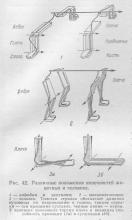
As a result of all these movements, the animal, rising above the ground, stands on all four legs and can use them when walking and running.
Man, the only one of all primates, walks in an upright position, relying only on his hind limbs, which he has become lower, located on the continuation of the vertical axis of the body.
Forelimbs , which became upper in a person due to his vertical position, lost their locomotor function. Thanks to labor activity, which singled out a person from the environment of animals, they turned into a kind of grasping organ, adapted to perform various and subtle movements necessary during work. The hand has become an organ of labor. Accordingly, the bones of the hand are thinner and lighter than the bones of the lower limb, and, in addition, are connected to each other by very mobile joints. In particular, pronation and supination are developed (rotation of the radii with a turn of the hand with the rear up and vice versa). In addition to the mobility of the joints, the freedom of movement of the upper limb depends to a large extent on the presence of the collarbone, which puts the limb to the periphery. Another feature in the skeleton of the upper limb, characteristic of man, is twisting, torsio, of the shoulder, which occurs in connection with the vertical position of the body; since the human chest is compressed from front to back, and not from the sides, as in four-legged animals, the human scapula is adjacent to the back surface of the chest, being turned with its articular fossa to the lateral side (in tetrapods, the fossa is turned downwards). Depending on this, the articular surface of the head of the shoulder, which articulates with the scapula, rotates inward by almost 90 ° in relation to the distal epiphysis of the same bone. Shoulder torsion develops gradually over individual development person.
The brush especially adapts to work. Carpal bones become small; on the contrary, the fingers lengthen and become very mobile. The thumb is set aside and can be opposed (opposed) to all other fingers, including V, which monkeys cannot do; some of them can bring the thumb no further than III. In addition, their thumbs are short. Thanks to this structure, the human hand is able not only to grasp an object, as is the case with great apes, but also to clasp it, which is of great importance for the “grasping function” of the hand during work. All these features of the structure of the upper limb of a person arose as a result of the improvement of the hand in the process of labor activity. Therefore, as Engels says, the hand is the organ of labor and, at the same time, its product.
lower limbs of a person serve only to move the body in space and at the same time are supports on which the entire weight of the body rests, therefore the bones of the lower limb are thicker, more massive and the mobility between them is much less than that of the upper limb.
The foot, as the ultimate support of the body, has lost the properties of the grasping leg found in monkeys, as a result of which the fingers, which do not play any role in the support, have been greatly shortened. The thumb is in line with the others and does not differ in particular mobility, as on the hand. The foot has acquired the shape of an arch, smoothing out, like a spring, shocks and shocks when walking and running.
The first rudiments of limbs in humans appear on the 3rd week of embryonic life in the form of horizontal protrusions on the sides of the body of the embryo, resembling fins. The protrusions expand into a roundish plate (rudiment of the hand and foot), in which fingers cannot yet be distinguished. The latter are outlined in the plate later in the form of five rays. Then the elements of the forearm and lower leg develop, and finally, the shoulder and thigh. Thus, the development of individual links of the limb proceeds in this order: first, the distal links, then the middle ones, and finally the proximal ones, as if from the body, during the development of the upper limb, first the hand, then the forearm and, finally, the shoulder, with the development of the lower - the foot, shin, thigh.