metacarpus, metacarpus, formed by five metacarpal bones, ossa metacarpalia, which, by type, belong to short tubular bones with one true epiphysis (monoepiphyseal bones) and are named in order I, II, III, etc., starting from the side of the thumb. Each metacarpal bone consists of a base, basis, diaphysis, or body, corpus, and a rounded head, caput. The bases of the II-V metacarpal bones have flat articular facets at their proximal ends for connection with the bones of the second row of the wrist, and on the sides for articulation with each other. Foundation I metacarpal bone has a saddle-shaped articular surface, attached to the os trapezium, while the lateral facets are absent. The base of the II metacarpal bone forms a notch in the form of an angle, covering the os trapezoideum; there is a tubercle on the ulnar side of the base of the fifth metacarpal bone. The heads of the metacarpal bones are convex articular surfaces for articulation with the proximal phalanges of the fingers. On the sides of the heads there are rough pits - the places of attachment of the ligaments. The shortest and at the same time the thickest of the metacarpal bones is I, relating to the thumb. Metacarpal II is the longest, followed by III, IV and V.
Finger bones
Finger bones, ossa digitorum minus, are small, short tubular bones lying one behind the other with one true epiphysis (monoepiphyseal bones), called phalanges. Each finger consists of three phalanges: proximal, phalanx proximalis, medium, phalanx media, and distal, phalanx distaiis. The exception is the thumb, which has only two phalanges - proximal and distal. In all animals, it is less developed than others and reaches its greatest development only in humans. The base of the proximal phalanx bears a single articular fossa for articulation with a round head. of the corresponding metacarpal bone, and the bases of the middle and distal phalanges have two flat fossae separated by a comb. They articulate with the heads of the proximal and middle phalanges, respectively, in the form of a block with a notch in the middle. The end of the phalanx is flattened and bears roughness, tuberositas phalangis distaiis. In the area of the metacarpophalangeal and interphalangeal joints of the hand, at the site of attachment of the tendons, there are sesamoid bones. They are constant on thumb and unstable on the rest.
shoulder blade
The scapula, scapula, is a thin, flat, triangular bone, which is freely laid between the muscles, articulating movably with the lateral section with the clavicle and humerus. Two surfaces are distinguished in the scapula: the anterior, costal, facies costalis, facing the ribs, and the dorsal, facies dorsalis, facing backwards. One of the edges, medial, margo medialis, with a freely hanging arm, is located almost parallel to the spine at the level from II-III to VII-VIII ribs. The second edge is the upper one, margo superior, facing upwards and has a notch, incisura scapulae, sometimes turning into a hole through which the suprascapular nerve passes. The third edge is the lateral, margo lateralis, the most thickened of all the edges, bifurcated along its entire length and descends into roughness on the costal and dorsal surface of the scapula. These three edges converge at the corners, of which the lower one, angulus inferior, is rounded and extended downward, the upper one, angulus superior, ACUTE, faces upward, lateral, angulus lateralis, is thickened, equipped with an articular cavity, cavitis glenoidalis, for articulation with the head humerus. The articular cavity is separated from the cervical scapula, collum scapulae. Above and below the articular cavity there are two tubercles: supraarticular, tuberculum supraglenoidale, to which the tendon of the long head of the biceps brachii is attached, and subarticular, tuberculum infraglenoidale, - the place of attachment of the tendon of the long head of the triceps brachii. Between the lateral angle and the notch, there is a coracoid process, processus coracoideus, of a curved shape, the initial section of which is directed upward and forward, the final section is forward and outward. This process, up to 13-15 years old, is connected to the scapula through cartilage.
The costal surface of the scapula is concave, especially in the upper lateral region, and is called the subscapular fossa, fossa subscapularis. The bundles of the subscapularis muscle begin in it. The dorsal surface of the scapula, facing backwards, is convex and is divided by the scapular spine, spina scapulae, into two pits: a smaller supraspinatus, fossa supraspinata, and a larger one, occupying approximately the lower ⅔ of the surface, infraspinatus, fossa infraspinata.
The scapular spine is more expanded from the lateral side, where it passes at an angle into the humeral process, acromion. At the apex of the process there is a small oval articular surface for connection with the acromial end of the clavicle.
Ossification. The scapula has three most permanent ossification points: in the body, the coracoid process and the lower angle with the adjacent part of the medial edge. In the body, the ossification point appears on the 2nd month of intrauterine development, in the coracoid process - on the first year of life, in the area of the lower angle and medial edge - on the 15-17th year. The coracoid process fuses with the body at the age of 14-16, the lower angle and medial edge - at the age of 21-25.
Collarbone
The clavicle, clavicula, is a curved tubular bone located between the sternum and the acromial process of the scapula. In the clavicle, the middle part and two ends are distinguished: the sternal, extremitas sternalis, facing the sternum, and the shoulder, extremitas acromialis, the acromial process of the scapula. The sternal end is more expanded and massive in comparison with the humeral end, it is provided with a saddle-shaped articular surface for connection with the sternum. The humeral end is thickened, carries an articular surface for connection with the humeral process. The upper surface of the clavicle is smooth, on the lower there is a conical tubercle, tuberculum conoideum, - the place of attachment of the ligaments. The part of the clavicle, located near the sternal end, is turned forward with a bulge; the department closest to the acromial end has a bulge directed backwards.
Ossification. The clavicle has two points of ossification: in the body and the epiphysis facing the sternum. The ossification point in the body appears first in the entire skeleton at the 6th month of intrauterine development, at the sternal end - at the 16-20th year, fusion with the body occurs at the 21st - 25th year of life.
BONES OF THE FREE UPPER LIMB
The free upper limb consists of three sections. The proximal section is the shoulder, the middle section is the forearm and the distal section is the hand. The skeleton of the shoulder forms the humerus, the bones of the forearm, ossa antebrachii, consist of the radius and ulna. The skeleton of the hand, manus, includes the bones of the wrist, metacarpus and phalanges of the fingers.
Brachial bone
The humerus, humerus, is a long tubular bone in which a body is distinguished - the diaphysis and two ends - the epiphyses: upper (proximal) and lower (distal). The upper end connects to the scapula, the lower end to the bones of the forearm. At the upper end of the humerus is the head, caput humeri, facing upward and medially, covered with hyaline cartilage and representing almost half of the ball. The head is separated from the rest of the upper end of the humerus by the anatomical neck, collum anatomicum. Behind it are two hillocks: a large one, tuberculum majus, facing outward, and a small one, tuberculum minus, forward. The tubercles continue downward into ridges: a large tubercle, crista tuberculi majoris, and a small one, crista tuberculi minoris. Between the crests and tubercles is the intertubercular groove, sulcus intertubercularis, which is the site of passage of the tendon of the long head of the biceps brachii.
The body of the humerus, corpus humeri, is cylindrical in the upper part and triangular in the lower part. Approximately in the middle of the anterior lateral surface of the body is the deltoid tuberosity, tuberositas deltoidea, - a trace of the attachment of the deltoid muscle. Behind the tuberosity on the posterior surface is the groove of the radial nerve, sulcus n. radialis, which spreads spirally from top to bottom from the medial edge to the lateral. At the point of transition of the upper end into the body is the surgical neck, collum chirurgicum, so named because bone fractures most often occur in this area. The lower end of the humerus - the condyle, condylus humeri, has a triangular shape with the base facing downwards. Its lateral sections form the medial and lateral epicondyles, epicondyli medialis et lateralis, which are the starting point for the muscles of the forearm and ligaments elbow joint. The medial epicondyle, larger in size, on the back side bears the groove of the ulnar nerve, sulcus n. ulnaris. On the basis of the lower end of the humerus are located: medially - the block of the humerus, trochlea humeri, - the articular surface for articulation with ulna, laterally - head, capitulum humeri, - articular surface for radius. On the back surface of the lower end, above the block, there is a fossa of the olecranon, fossa olecrani, where the process enters ulna. Two fossae lie on the anterior surface: coronal, fossa coronoidea, and radial, fossa radialis.
Ossification. The humerus has seven ossification points, of which one appears in the body (7-8th week of intrauterine development), three in the upper (proximal) epiphysis, and three in the lower (distal). In the upper epiphysis, they occur sequentially in the region of the head, in the region of the greater and then the lesser tubercle (2-5 years), in the lower epiphysis - in both padcondyles and the block (8-12 years). At the age of 18-20, these islands of ossification merge.
Forearm bones
The ulna is located on the medial side of the forearm, the radius - on the lateral. Both bones are long tubular bones, in which the upper proximal and lower distal ends, or epiphyses, are distinguished, and the body is the diaphysis. The ends of the radius and ulna are located on different levels. In the proximal section, the upper end of the ulna is located above, in the distal section, the lower epiphysis of the radius occupies a lower position. The ends of the bones are connected to each other by means of joints. In the rest of the length between the bones there is a connective tissue interosseous membrane.
Elbow bone
The proximal end of the ulna, ulna, is massive, widened. It connects to the block of the humerus. It has two processes: the upper - ulnar, olecranon, and the lower - coronal, processus coronoideus, which limit the block-shaped notch, incisura thochlearis, open anteriorly. The olecranon is well palpable under the skin, the coronoid is covered by the muscles located around the elbow joint. For articulation with the head of the radius, on the lateral side of the coronoid process there is a radial notch, incisura radialis. Below the notch is the crest of the supinator muscle for attaching the muscle of the same name. Tuberosity, tuberositas ulnae, is determined in front and below the coronoid process, for attaching the tendon shoulder muscle.
The body of the ulna is trihedral and has anterior, posterior, and medial surfaces separated from each other by edges. The distal end is much smaller than the upper one, bears the head, from the medial side of which the styloid process, processus styloideus, departs, well palpable under the skin. On the lateral surface of the head is the articular circle, circumferentia articularis, which articulates with the ulnar notch of the radius. The ulna is easily palpable under the skin from behind all the way from the ulna to the styloid process. In front, the bone is covered with muscles and tendons.
Radius
The upper proximal end of the radius, radius, forms a head, caput radii, equipped at the top with a flat fossa for articulation with the head of the humerus. On the lateral surface of the head there is an articular circle, circumferentia articularis, for articulation with the notch of the ulna. Slightly below the head, the radius forms a neck, collum radii, below and medially to which there is a tuberosity, tuberositas radii, for attaching the tendon of the biceps muscle.
The body of the radius is curved, with a bulge facing laterally. It has three surfaces, anterior, posterior and lateral, separated by three edges. The lower, distal end of the radius is thickened, forms the styloid process, processus slyloideus, on the lateral side, and the ulnar notch, incisura ulnaris, for the head of the ulna on the medial side. From below, the distal epiphysis has a carpal articular surface, facies articularis carpea, covered with cartilage, for articulation with the bones of the proximal row of the wrist. In the radius, you can feel the head and its entire lower section with the styloid process under the skin.
Ossification. Five ossification points appear in each of the bones of the forearm: one in the body (end of the 2nd month of fetal development) and two in the epiphyses (2-19 years). Initially, ossification points appear in the radius (2-5 years), then in the ulna (5-19 years), and the lower epiphyses are the first to undergo ossification. The fusion of the epiphyses with the body occurs in the reverse order: first, the upper epiphyses of the ulna (14 years old) grow, then the upper epiphyses of the radius (18-19 years old) and, at the age of 21, the lower epiphyses of both bones.
Hand bones
Hand bones, ossa manus include the bones of the wrist, metacarpus and phalanges of the fingers.
wrist bones
Wrist, carpus, consists of 8 small short bones, ossa carpi, located in two rows: proximal and distal. The bones of the wrist have a variety of sizes and shapes, which is reflected in their names. The composition of the proximal row of bones of the wrist (counting from the side of the thumb) includes: the scaphoid, os scaphoideum, lunate, os lunatum, trihedral, os triquetrum, and pisiform, os pisiforme. The pisiform bone is one of the sesamoid bones embedded in the tendon of the muscle. The bones of the proximal row (except for the pisiform), connecting with each other, form an arc, the convex surface of which is connected to radius, concave - with the bones of the distal row of carpal bones. The distal row consists of: trapezoid bone, os trapezium, trapezoid, os trapezoideum, capitate, os capitatum, and hooked, os humatum, bones. top surface the bones of this row are covered by an arc formed by the bones of the proximal row, the lower surface, which has a stepped character, is connected to the bones of the wrist.
The bones of the wrist have articular surfaces to connect with each other and adjacent bones. On the scaphoid and trapezoid bones on the palmar side there are tubercles, and on the hooked bone there is a hook, hamulus, which serve to attach tendons and ligaments. The bones of the wrist are located so that on the palmar side the wrist is concave in the form of a groove or groove, on the back it is convex. The groove of the wrist, sulcus carpi, is limited on the medial side by the pisiform bone and the hook of the hamate bone, and on the lateral side by the tubercles of the trapezoid bone and the navicular bone. It serves for the passage of tendons, blood vessels and nerves. It is difficult to feel the bones of the wrist separately. The pisiform bone, which is located under the skin in the upper medial angle of the palm, is most easily palpated and is displaced during palpation. On the palmar side, during flexion and extension of the hand, a tubercle of the bone is felt - the trapezium, on the back side - the capitate and trihedral bones.
Ossification. The bones of the wrist have one ossification point in each of them. The first to appear are ossification points in the capitate and hooked bones (in the 1st year of life), the last in the pisiform (at 12-13 years).
Metacarpal bones
The metacarpus, metacarpus, consists of 5 metacarpal bones, ossa metacarpalia, tubular in shape. The name of each of them corresponds to their serial number, counting from the thumb (I-V). The metacarpal bone has a body and two ends. The body of the metacarpal bones is irregularly triangular in shape, concave on the palmar side. The proximal end - the base, basis, is connected to the second row of metacarpal bones, and the distal end - the head, caput, - to the proximal phalanx. The articular surfaces of the bases of II - IV metacarpal bones are flat, saddle-shaped. On the basis of the III metacarpal bone there is a process that protrudes between the capitate and trapezoid bones. The articular surface on the head is convex, on its lateral sections there are roughnesses for attaching ligaments. The shortest is the I metacarpal bone, the longest is the III.
Phalanges of fingers
Each finger consists of phalanges, phalanges digitorum manus. The first finger has two phalanges - proximal and distal, the rest have three - proximal, middle and distal. The largest sizes are in the proximal phalanges, the smallest in the distal ones. Each phalanx is a tubular bone in shape and has a body, corpus phalangis, flattened from front to back, and two ends: the proximal one is the base, basis phalangis, and the distal one is the head, caput phalangis. The heads of the first and second phalanges are block-shaped, and the third - tuberosity, tuberositas phalangis distalis. The bases of the first phalanges bear articular surfaces in the form of pits for articulation with the heads of the metacarpal bones. On the bases of the second and third phalanges, the articular surface corresponds to the block-like surface of the heads of the first two phalanges that articulates with it and has a guiding ridge. Sesamoid bones are located at the level of the joints between the metacarpal bones and the proximal phalanges of I, less often V and II fingers on the palmar side.
Ossification. The bones of the metacarpus and phalanges of the fingers have two ossification points each - in the body and in one of the epiphyses. The ossification point in the body occurs at the 2-3rd month of intrauterine development, in the epiphysis at 3-10 years of age, and in the II-V metacarpal bones, the ossification centers are located in the head, and in the I metacarpal bone and in all phalanges - in the bases . Fusion of the body with the epiphysis occurs at the age of 18-21. Sesamoid bones of the first finger receive ossification points at the age of 12-16.
human limbs
joints shoulder girdle provide freedom of movement of the hands of a person. Bundles upper limbs provide conditions for mobility restrictions within the framework of anatomical features. We offer you a material that describes in detail the anatomical structure of the upper shoulder girdle, its functional features. All joints of the human upper extremities are considered.
The joints of the girdle of the upper extremities (shoulder girdle) connect the clavicle to the sternum and to the scapula, forming the sternoclavicular and acromioclavicular joints.
sternoclavicular joint
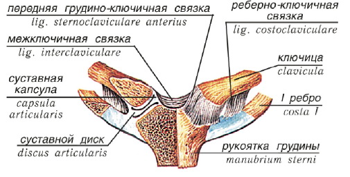
The sternoclavicular joint is flat, multiaxial, formed by the sternal articular surface of the clavicle and the clavicular notch of the sternum handle. The shape of the articular surfaces of the bones articulating in it approaches the saddle. Between the articular surfaces is the articular disc, which fuses with the capsule along the periphery and divides the articular cavity into two chambers. The thin joint capsule is strengthened by the anterior and posterior sternoclavicular ligaments, which are woven into the fibrous membrane of the joint capsule in front and behind. There is also an interclavicular ligament connecting the sternal ends of both clavicles, and a costoclavicular ligament located laterally at some distance from the joint.
 The sternoclavicular joint is triaxial. The range of motion is limited in the joint. According to the three axes in the joint, forward and backward movements, raising and lowering, as well as some rotation can be performed. In addition, it is possible Roundabout Circulation, in which the acromial end of the clavicle describes an ellipse.
The bones of the fingers (ossa digitorum manus) - phalanges are short tubular bones. Each finger has three phalanges located one behind the other: proximal, middle and distal. The exception is the thumb, which has only two phalanges - proximal and distal. The base of the proximal phalanx articulates with the head of the corresponding metacarpal bone, and the bases of the middle and distal phalanges articulate with the heads of the proximal and middle phalanges, respectively. The distal phalanges are also called nail.
CONNECTIONS OF THE BONES OF THE FOREARM WITH THE HAND
wrist joint (articulatio radiocarpea) is formed by the lower articular surface on the distal epiphysis of the radius and the articular surfaces of the first row of carpal bones (scaphoid, lunate and triangular). The ulna is shorter than the radius, therefore, at its distal end there is an articular disc, which fuses with the radius and together with it forms the articular cavity of the wrist joint.
The bones involved in the formation of the wrist joint are interconnected by interosseous ligaments. The capsule of the wrist joint is strengthened by the radial and ulnar collateral ligaments located on the lateral surfaces of the joint. On the palmar side of the wrist joint is the palmar radiocarpal ligament. On the back side, the capsule of the radiocarpal joint is supported by the dorsal radiocarpal ligament.
By the number of bones involved, the joint is complex, and by the shape of the articular surfaces it belongs to ellipsoidal with two axes of rotation. Around the sagittal axis, the hand is abducted and adducted in the wrist joint; around the frontal axis - flexion and extension.
JOINING THE BONES OF THE HAND.
Mid-carpal articulation(articulatio mediocarpea) is formed between the first and second row of carpal bones. The pisiform bone does not participate in this connection, since it is sesamoid. The joint has its own capsule. The ligaments that strengthen the joint are the same as in the wrist. The joint is elliptical in shape, biaxial. Here, circular movements with a brush are also possible.
Intercarpal joints(articulationes intercarpea) unite individual bones of the wrist. They are reinforced by interosseous carpal ligaments stretching between articular surfaces facing each other.
Movements in the joints of the hand are performed around two mutually perpendicular axes: around the frontal - flexion and extension; around the sagittal - abduction and adduction. The intercarpal joints are stiff joints that form the solid base of the hand, providing a variety of finger movements.
The flexor retinaculum is not directly related to the joints of the hand, it is thrown in the form of a bridge from the carpal radial eminence to the carpal ulnar eminence through the carpal groove, turning the latter into a carpal tunnel. In the channel pass median nerve, as well as the tendons of the flexors of the fingers, hence the name of the ligament - the flexor retinaculum.
Carpometacarpal joints(articulationes carpometacarpeae) are formed by the second row of carpal bones adjacent to the bases of the metacarpal bones. With the exception of the carpometacarpal joint of the thumb, all these joints are flat, strengthened both from the back and from the side of the palm by tightly stretched ligaments, as a result of which mobility in them is extremely insignificant. They can only slide in one direction or another. They belong to tight joints that strengthen the hand and increase the resistance of the palm during forceful movements of the muscles - the flexors of the fingers.
The carpometacarpal joint of the little finger has a slightly greater mobility. Due to the fact that the articular surface of the base of the fifth metacarpal bone is almost saddle-shaped, the little finger can be very limitedly opposed to the thumb. The common cavity of the carpometacarpal joints, surrounded by a capsule, has the shape of a transverse slit, which communicates with the midcarpal joint and the intermetacarpal joints.
Metacarpal joints(articulationes intermetacarpeae) are located between the bases of the last four metacarpal bones adjacent to each other; in depth, these connections are strengthened by strong interosseous ligaments. The capsules of the intermetacarpal joints are supported by transverse dorsal and palmar ligaments.
Carpometacarpal joint of thumb completely isolated from the rest of the carpometacarpal joints and differs sharply from them in its structure and mobility. It is formed by the saddle-shaped articular surfaces of the trapezium and the base of the first metacarpal bone, articulated with each other, surrounded by a wide articular capsule. Being a saddle joint, movements are allowed in the joint around two axes: transverse and sagittal. Around the first of them, flexion and extension of the thumb occurs along with its metacarpal bone, but since the axis is not completely transverse, the finger, when bent, shifts towards the palm, opposing the little finger and the rest of the fingers. Movements around the sagittal axis consist of abduction and adduction of the thumb to the index finger. In addition to the movements described, there is also a rotational movement of the thumb.
Metacarpophalangeal joints(articulationes metacarpophalangeae) are formed by the heads of the metacarpal bones and pits on the basis of the proximal phalanges, they are spherical in shape. The ligamentous apparatus consists of two auxiliary lateral ligaments. Between the heads of the metacarpal bones from II to V on the palmar side stretch the transverse metacarpal ligaments. Movements in the metacarpophalangeal joints are performed around two axes: frontal - flexion and extension, and sagittal - abduction and adduction of the finger. In addition, in these joints, the finger can also make a circular motion.
Interphalangeal joints(articulationes interphalangeae manus) are located between the head and the base of adjacent phalanges of the finger, they are block-like joints that allow flexion and extension around the frontal axis. Auxiliary ligaments run along the lateral surfaces of the joint.
SKELETON OF THE LOWER LIMB AND ITS JOINTS.
The skeleton of the lower limb consists of a belt and a free lower limb.
LOWER LIMB BELT
The belt of the lower limb (cingulum membri inferioris), in contrast to the belt of the upper limb, is a closed bone ring formed by a pair pelvic bone and sacrum (described above). Such a structure of the lower limb belt provides good support for the entire body and head, and the movement of its free sections.
The pelvic bone has a complex structure, refers to flat bones and is formed by three fused bones: the ilium, pubic and ischium. Until the age of 16, the bones are connected to each other by cartilage, then they grow together around the acetabulum, which is the articular fossa in the hip joint.
ILIUM
The ilium (os ilium) has a body adjacent to the acetabulum and a wing located upward from it. The inner surface of the wing is concave and smooth and is called the iliac fossa. The relief of the outer surface of the bone is due to the attachment of muscles. The upper free edge of the wing is called the iliac crest.
In front and behind the crest ends, respectively, with the anterior and posterior superior iliac spines, under which the anterior and posterior inferior awns are located. The wing is separated from the body of the ilium by an arcuate line. At the junction of the ilium with the pubis, there is an iliopubic eminence. In the back of the wing there is an ear-shaped articular surface, which is involved in the formation of the sacroiliac joint. Behind and above the ear-shaped surface is the iliac tuberosity - the place of attachment of the ligaments.
PUBIC BONE
The pubic bone (os pubis) is located anterior and medial to the acetabulum. It has a short thickened body adjacent to the acetabulum, the upper and lower branches located at an angle to each other. At the junction of the branches, an articular surface is formed, which articulates with the similar surface of the pubic bone of the other side. On the lower surface of the superior branch of the pubic bone there is an obturator groove.
ISCHIUM
The ischium (os ischii) is located behind and below the acetabulum. It has a body adjacent to the acetabulum and a branch located at an angle to the body. The angle is thickened and is called the ischial tuberosity. Behind the ischial tubercle is the ischial spine, which separates the small ischial notch from the large one.
The branch of the ischium fuses with the lower branch of the pubic bone. The branches of the pubic and ischial bones surround the obturator foramen, closed by the membrane of the same name. The membrane, being thrown through the obturator sulcus, turns it into the eponymous canal through which the vessels and nerve pass.
PELVIC JOINTS
The pelvic bones, connecting with each other and the sacrum, form the pelvis. At the junction of both pubic bones is the symphysis - a semi-movable joint. At the junction of the pelvic bones with the sacrum, a stiff joint is formed, where strength is combined with mobility. In connection with upright posture, the human pelvis is a support for the viscera and a place for transferring weight from the trunk to the lower limbs, as a result of which it experiences a huge load.
sacroiliac joint(articulation sacroiliaca) is formed by flat ear-shaped articular surfaces of the sacrum and ilium. It is strengthened by the anterior and posterior sacroiliac ligaments, as well as the interosseous ligaments, which are the strongest ligaments in the human body. As noted above, the joint is stiff, flat in shape, multiaxial in function, but there are practically no movements in it.
The sacrum is connected to the pelvic bone by two ligaments: the sacrotuberous - with the ischial tuberosity and the sacrospinous - with the ischial spine.
The described ligaments complement the bony walls of the pelvis in its posterior-lower section and turn the large and small ischial notches into the large and small openings of the same name.
Pubic symphysis(symphysis pubica) or semi-joint is formed between two pubic bones. The articular surfaces of the pubic bones are covered with hyaline cartilage. Between them there is a fibrocartilaginous plate, in which a narrow joint space is formed. The role of the articular capsule here is performed by the perichondrium. The pubic symphysis is supported by the superior and inferior pubic ligaments. Under the latter, a subpubic angle is formed. In this connection, small displacements of the bones relative to each other are possible due to the elasticity of the cartilage.
pelvis as a whole
Pelvis (pelvis) in the form of a bone ring formed by the connection of the pelvic bones with each other and with the sacrum, serves to connect the body with free lower limbs. It is divided into 2 sections: the upper, wide - the large pelvis and the lower, narrow - the small pelvis.
Large pelvis is a part abdominal cavity, limited laterally by deployed wings ilium. In front, it has no bony walls, and behind it is limited by the lumbar vertebrae.
The large pelvis is separated from the small pelvis by a border line formed by the cape, the arcuately curved edge of the ilium, the upper branches of the pubic bones and the upper edge of the pubic symphysis. The opening thus limited is called the entrance or the upper aperture of the small pelvis, below which lies its cavity.
In front, the wall of the small pelvis is short and is formed by the pubic bones and their joints, as well as the obturator membranes. Behind the wall is long and consists of the sacrum and coccyx. The lateral walls of the small pelvis are formed by the bodies of three bones adjacent to the acetabulum, branches of the pubic and ischial bones, as well as ligaments that run from the sacrum to the ischial tuberosities and awns. The exit from the cavity of the small pelvis or the lower aperture is closed by the muscles and fascia of the perineum.
Sexual differences begin to appear at the onset of puberty. Bones female pelvis smoother and thinner. The wings of the ilium in women are deployed to the sides. The upper aperture of the small female pelvis has a transverse-oval shape, while in the male pelvis it is longitudinally oval. The cape of the male pelvis protrudes more forward. The pelvic surface of the sacrum is more concave, while in women, on the contrary, the sacrum is relatively wider and at the same time more flat. The exit from the small pelvis in men is much narrower than in women. The ischial tubercles, which limit the lower aperture in women, are further apart, and the coccyx protrudes less forward. The subpubic angle in women is obtuse and arch-shaped, while in men it is more acute. The pelvic cavity in men is funnel-shaped, in women the pelvis is cylindrical.
The bones of the fingers, ossa digitorum minus, are small, short tubular bones lying one behind the other with one true epiphysis (monoepiphyseal bones), called phalanges. Each finger consists of three phalanges: proximal, phalanx proximalis, middle, phalanx media, and distal, phalanx distaiis. The exception is the thumb, which has only two phalanges - proximal and distal. In all animals, it is less developed than others and reaches its greatest development only in humans. The base of the proximal phalanx bears a single articular fossa for articulation with a round head. of the corresponding metacarpal bone, and the bases of the middle and distal phalanges have two flat fossae separated by a comb. They articulate with the heads of the proximal and middle phalanges, respectively, in the form of a block with a notch in the middle. The end of the phalanx is flattened and bears a roughness, tuberositas phalangis distaiis. In the area of the metacarpophalangeal and interphalangeal joints of the hand, at the site of attachment of the tendons, there are sesamoid bones. They are constant on the thumb and inconsistent on the rest.
Wrist joint, art. radiocarpea. In most mammals, it is block-shaped. As the ability to pronate and supinate is acquired, a separate joint develops between the ray and the ulna - the distal radioulnar, art. radioulnaris distalis, which, together with the proximal radioulnar joint, forms a single combined articulation with a vertical axis of rotation. In this combined articulation, the radius moves around the ulna, as a result of which the distal epiphysis of the radius increases significantly. On the contrary, the distal epiphysis of the ulna lags behind in its development and becomes shorter than the beam, but a special articular disc, discus articularis, appears on it. In humans, due to the largest volume of supination and pronation, discus articularis reaches its highest development and takes the form of a triangular fibrocartilaginous plate, fibrocartilago triangulare, which fuses with the distal epiphysis of the radius and together with it forms the articular cavity of the proximal section of the wrist joint. Consequently, the ulna participates in the wrist joint only through the named cartilaginous disk, not having a direct relation to this articulation, therefore the proximal part of this joint is called not the forearm joint, but the wrist joint.
In accordance with the above articular cavity art. radiocarpea is formed by facies articularis carpea of the ray and discus triangularis, and the articular head of this joint is formed by the proximal surface of the first row of carpal bones, os scaphoideum, lunatum et triquetrum, which are interconnected by interosseous ligaments, ligg. intercarpea. According to the number of bones involved, the joint is complex, and according to the shape of the articular surfaces, it belongs to ellipsoidal ones with two axes of rotation (sagittal and frontal).
1. Mid-carpal articulation, art. mediocdrpea, is located between the first and second row of carpal bones, minus the pisiform bone, which is sesamoid. The articular cavity of this joint is the distal surface of the first row of carpal bones. The proximal surface of the second row of the wrist forms the articular head.
Both carpal joints (wrist and mid-carpal) have independent articular capsules attached along the edges of their articular surfaces. To strengthen the capsule of the wrist joint, auxiliary ligaments serve from the radial and ulnar sides: lig. collaterale carpi radiate, running from the styloid process of the ray to the scaphoid, and lig. collaterale carpi ulnare, extending from the styloid process of the ulna to os triquetrum and os pisiforme. On the palmar side of the wrist joint is lig. radiocarpeum palmare, which, starting widely from the styloid process and from the edge of the articular surface of the beam, is attached to os scaphoideum, lunatum, triquetrum et capitatum in several bundles. On the back side, the capsule of the wrist joint is supported by lig. radiocarpeum dorsale, which runs from the ray to the bones of the first row of carpal bones. At the site of attachment of the ligaments of the wrist joint to the bones, the latter include blood vessels and nerves, damage to which during operations entails pathological changes in the bones. Capsule art. mediocarpea also captures the last four carpometacarpal joints that communicate with each other. Except art. mediocarpea, separate bones of the wrist, connected to each other by interosseous ligaments, ligg. intercarpea interossea, in some places are articulated with each other by articular surfaces facing each other. Such joints are called intercarpal, articulationes intercarpeae.
The intercarpal joints are supported by a series of short ligaments, running mostly transversely from one bone to another on the back, ligg. intercarpea dorsalia, and palmar, ligg. intercarpea palmaria, sides. On the palmar side, in addition, note bundles diverging from the capitate to neighboring bones, lig. carpi radiatum. Movements in the wrist joints are performed around two mutually perpendicular axes passing through the head of the capitate, around the frontal (flexion and extension) and around the sagittal (abduction and adduction). These movements are inhibited by ligaments that are located perpendicular to the axes of rotation and at their ends, namely: collateral - at the ends of the frontal axis, dorsal and palmar - at the ends of the sagittal axis. Therefore, the former inhibit abduction and adduction around the sagittal axis, while the latter inhibit flexion and extension around the frontal axis. As in all biaxial joints, circumductio is also possible here, in which the ends of the fingers describe a circle.
The wrist joint is powered by the rete ariticulare formed by the branches of a. radialis, a. ulnaris, aa. interosseae anterior et posterior. Venous outflow occurs in the veins of the same name, carrying blood into the deep veins of the forearm - vv. ulnares, vv. radiales, vv. interosseae. The outflow of lymph is carried out through the deep lymphatic vessels in the nodi lymphatici cubitales. Innervation - from p. radialis, n. ulnaris, n. medianus.
2. Articulation of the pisiform bone, art. ossis pisiformis, represents a separate joint in which the pisiform bone articulates with the os triquetrum. Two ligaments come from the pisiform bone: lig. pisohamatum to the hamate and lig. pisometacarpeum to the base of III-V metacarpal bones. These ligaments are a continuation of the tendon m. flexor carpi ulnaris, in the thickness of which the named sesamoid bone is laid.
3. The flexor retinaculum, retinaculum flexorum, is not directly related to the joints of the hand; it spreads in the form of a bridge from eminentia carpi radialis to eminentia carpi ulnaris through the carpal groove, sulcus carpi, turning the latter into a canal, canalis carpi. P. medianus, as well as tendons of the flexors of the fingers, pass through the canal, hence the name of the ligament - retinaculum flexorum.
4. Carpometacarpal joints, artt. carpometacarpeae, are formed by the second row of carpal bones and the bases of the metacarpal bones. With the exception of the carpometacarpal joint of the thumb, all these joints are flat, strengthened both from the rear and from the side of the palm by tightly stretched ligaments, ligg. carpometacarpea dorsalia et palmaria, as a result of which the mobility in them is extremely insignificant. They can slip 5-10 ° in one direction or another. They belong to the category of tight joints, strengthening the root section of the hand and increasing the resistance of the palm during forceful movements of the multi-joint muscles - finger flexors.
The carpometacarpal articulation of the little finger has somewhat greater mobility. Due to the fact that the articular surface of the base of the fifth metacarpal bone is almost saddle-shaped, the little finger can be opposed to the thumb in a very limited size. The common cavity of the carpometacarpal joints, surrounded by a capsule, has the shape of a transverse slit, which communicates with the midcarpal joint and the intermetacarpal joints. The mentioned intermetacarpal joints, artt. intermetacarpeae, located between the bases of the last 4 metacarpals adjacent to each other; in depth, the articulating surfaces of the bases of these bones are connected by strong ligaments, ligg. metacarpea interossea. The capsules of the intermetacarpal joints are supported by transverse dorsal and palmar ligaments, ligg. metacarpea dorsalia et palmaria.
Carpometacarpal joint of the thumb, art. carpometacarpea pollicis, is completely isolated from the rest of the carpometacarpal joints and differs sharply from them in its structure and mobility. It is formed by the saddle-shaped articular surfaces of the os trapezium and the base of the first metacarpal bone that articulate with each other, surrounded by a wide articular capsule. Being a typical saddle joint, this joint allows movement around two mutually perpendicular axes: the transverse one, passing through the os trapezium, and the sagittal one, going through the base of the first metacarpal bone. Around the first of the axes, the thumb is flexed and extended along with its metacarpal bone, but since the axis is not completely transverse, the finger, when flexed, shifts towards the palm, opposing the little finger and the rest of the fingers. This movement is called opposition, oppositio; the reverse movement is called repos "itio. Movements around the sagittal axis consist of abduction and adduction of the thumb to the index finger. The range of mobility is 45 - 60 ° for abduction and adduction and 35 - 40 ° for opposition and reverse movement. In addition to the movements described, there is also circum-ductio The saddle joint of the first finger progresses in the process of human evolution in connection with his labor activity.Thus, in Neanderthals this joint was apparently flattened and therefore performed less extensive movements than in modern man.
5. Metacarpophalangeal joints, artt. metacarpophalangeae, between the convex heads of the metacarpal bones and the pits at the base of the proximal phalanges, are close to ellipsoid in shape. The ligamentous apparatus consists of two auxiliary ligaments, ligg. collateralia, extending from the pits on the radial and ulnar surfaces of the metacarpal heads obliquely to the sides of the base of the proximal phalanges. On the palmar side there is a thickening containing fibrous cartilage, lig. palmare. In connection with this thickening between the heads of the metacarpal bones from II to V, transversely strong fibrous ligaments, ligg. metacarpea transversa profunda. Movements in the metacarpophalangeal joints are performed around two axes: frontal - flexion and extension of the entire finger with a range of motion of 90-100 ° and sagittal - abduction and adduction of the finger by 45 - 50 °. The last kind of movement is possible only with unbent fingers, when ligg. collateralia relaxed; when bent, they stretch and prevent lateral movements. In addition to the indicated movements, the finger can also make a circular motion, circumductio, in a rather extensive size.
6. Interphalangeal joints, artt. interphalangeae manus, located between the head and base of adjacent phalanges, are typical block-like joints that allow flexion and extension around the transverse (frontal) axis. Auxiliary ligaments, ligg. collateralia, go, on the sides of the joint
BONES OF THE BRUSH
The bones of the hand are divided into the bones of the wrist, metacarpus and the bones that make up the fingers, the so-called phalanges.
Wrist
Wrist, carpus, represents a collection of 8 short spongy bones- ossa carpi, arranged in two rows, each of which consists of 4 bones.
The proximal, or first, row of the wrist closest to the forearm is formed, counting from the thumb, by the following bones: scaphoid, os scaphoideum, lunate, os lunatum, trihedral, os triquetrum, and pisiform, os pisiforme. The first three bones, connecting, form an elliptical, convex articular surface towards the forearm, which serves for articulation with the distal end of the radius. The pisiform bone does not participate in this articulation, joining separately to the trihedral. The pisiform bone is a sesamoid bone that developed in the tendon m. flexor carpi ulnaris.
The distal, or second, row of the wrist consists of bones: a trapezoid, or a large polygonal, os trapezium, s. os multangulum majus, trapezoid, or small polygonal, os trapezoideum, s. os multangulum minus (BNA), capitate, os capitatum, and hook-shaped, os hamatum. The names of the bones reflect their shape. On the surfaces of each bone there are articular facets for articulation with neighboring bones. In addition, on the palmar surface of some bones of the wrist, tubercles protrude for attaching muscles and ligaments, namely: on the navicular bone - tuberculum ossis scaphoidei, on the os trapezium - tuberculum ossis trapezii and on the hamate bone - a hook, hamulus ossis hamati, which is why she received its name. The bones of the wrist in their totality represent a kind of arch, convex on the back and trough-like concave on the palmar. On the radial side, the carpal groove, sulcus carpi, is bounded by an eminence, eminentia carpi radialis, formed by the tubercles of the navicular bone and os trapezium, and on the ulnar side, by another eminence, eminentia carpi ulnaris, consisting of hamulus ossis hamati and os pisiforme.
In the process of human evolution in connection with his labor activity, the bones of the wrist progress in their development. So, in Neanderthals, the length of the capitate was 20-25 mm, and in modern man it increased to 28 mm. There is also a strengthening of the wrist region, which is relatively weak in great apes and Neanderthals. In modern man, the bones of the wrist are so firmly fastened by ligaments that their mobility decreases, but their strength increases. Therefore, the blow to one of the carpal bones is evenly distributed among the others and is weakened, as a result of which fractures in the wrist are relatively rare.
metacarpus
Metacarpus, metacarpus, is formed by five metacarpal bones, ossa tetacarpalia, which by type belong to short tubular bones with one true epiphysis (monoepiphyseal bones) and are called in order I, II, III, etc., starting from the side of the thumb. Each metacarpal bone consists of a base, basis, diaphysis, or body, corpus, and a rounded head, caput. The bases of the II-V metacarpal bones have flat articular facets at their proximal ends for connection with the bones of the second row of the wrist, and on the sides for articulation with each other. The base of the I metacarpal bone has a saddle-shaped articular surface, which is attached to the os trapezium, while the lateral facets are absent. The base of the II metacarpal bone forms a notch in the form of an angle, covering the os trapezoideum; on the ulnar side of the base of the V metacarpal bone there is a tubercle, tuberositas ossis metacarpi V. The heads of the metacarpal bones bear convex articular surfaces for articulation with the proximal phalanges of the fingers. On the sides of the heads there are rough pits - the places of attachment of the ligaments. The shortest and at the same time the thickest of the metacarpal bones is I, relating to the thumb. Metacarpal II is the longest, followed by III, IV and V.
Finger bones
Bones of fingers, ossa digitorum manus, are small, short tubular bones lying one behind the other with one true epiphysis (monoepiphyseal bones), called phalanges. Each finger consists of three phalanges: proximal, phalanx proximalis, middle, phalanx media, and distal, phalanx disialis, or nail. The exception is the thumb, which has only two phalanges - proximal and distal. In all animals, it is less developed than others and reaches its greatest development only in humans. The base of the proximal phalanx bears a single articular fossa for articulation with the round head of the corresponding metacarpal bone, and the bases of the middle and distal phalanges each have two flat fossae separated by a comb. They articulate with the heads of the proximal and middle phalanges, respectively, in the form of a block with a notch in the middle. The distal end of the nail phalanx is flattened and bears a roughness, tuberositas phaldngis distalis. In the metacarpophalangeal and interphalangeal joints of the hand, there are sesamoid bones, studied in detail by S. N. Kasatkin. They are constant on the thumb and inconsistent on the rest.
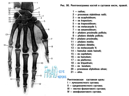
Ossification. The hand is the most convenient object for X-ray examination of the development of the skeletal system of a living person. On the radiograph of the hand of a newborn, it can be seen that only the diaphyses of tubular bones, which developed from the main ossification points in uterine life (starting from the 2nd month), underwent ossification. The epiphyses of the tubular bones and the bones of the wrist are still at the cartilaginous stage of development and therefore they are not visible on the radiograph. In the future, the following age-related changes in the skeleton of the hand are found:
1. Consistent appearance of ossification points in the bones of the wrist and in the epiphyses of tubular bones.
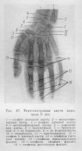
For easier memorization of the timing and order of ossification of the bones of the wrist, you can use the following technique: if you hold the radiograph in front of you with your fingers down and the radial edge to the right, then the order in which the ossification points appear in the bones of the wrist will correspond to the course of the hour hand, starting from the capitate bone. At the same time, it should be taken into account that the period of appearance of the bone core of a trihedral bone corresponds to the number of its faces (3 years); in the future, it is enough to add one year to each neighboring (clockwise) to get the ossification period. As a result, the order of ossification of the carpal bones will be as follows: capitatum (2 months), hamatum (3 months), triquetrum (3 years), lunatum (4 years), scapboideum (5 years), trapezium et trapezoideum (5 and 6 years) (Fig. 46, 47).
If the bone nuclei of the capitate and hamate bones are found on the radiograph of the newborn, then this, along with other symptoms, can serve as a sign of the full-term fetus. The ossification nuclei in the true epiphyses of short tubular bones appear in the 2-3rd year. At the opposite ends of these bones, independent ossification of false epiphyses (pseudoepiphyses) is sometimes traced. In the distal epiphyses of the long tubular bones, the ossification nuclei appear in the radius in the 1st or 2nd year and in the ulna in the 7th or 8th year. In sesamoid bones, ossification points appear in the prepubertal period: in the pisiform - in girls at 7-12 years old, in boys at 10-15 years old; in the metacarpophalangeal of the first finger - in girls at 10-15 years old, in boys at 13-17 years old. Sometimes sesamoid bones develop from two ossification centers that remain separate. These are the so-called ossa sesamoidea tripartita.
II. The onset of synostosis tubular bones in men at 19-23 years old, in women - at 17-21 years old. Knowledge of the timing and order of ossification makes it possible to determine various diseases endocrine glands and other body systems, when there is a perversion of ossification.
III. The aging of the hand skeleton is characterized by common signs of aging of the skeletal system.
It can be seen from the above that the skeleton of the hand, consisting of a large number of bones, undergoes significant age-related changes. Therefore, X-ray examination shows many morphological details that serve as reference points for determining the "bone" age.